Anti-Talin 1抗体
Anti-Talin 1 antibody
- BOND RX™ Validated
- KO Validated
- 了解详情
4
(4 Reviews)
|
(44 Publications)
Knockout Tested Rabbit Polyclonal Talin 1 antibody. Suitable for ICC/IF, WB, IHC-P and reacts with Mouse, Human samples. Cited in 44 publications.
查看别名
KIAA1027, TLN, TLN1, Talin-1
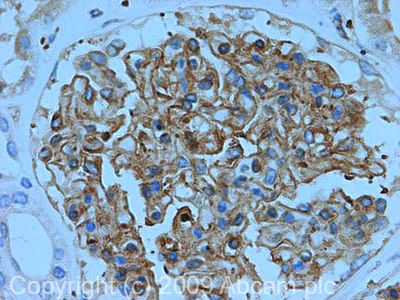
- IHC-P
Unknown
Immunohistochemistry (Formalin/PFA-fixed paraffin-embedded sections) - Anti-Talin 1 antibody (AB71333)
IHC image of Talin 1 staining in Human Normal Kidney FFPE section, performed on a BondTM system using the standard protocol F. The section was pre-treated using heat mediated antigen retrieval with sodium citrate buffer (pH6, epitope retrieval solution 1) for 20 mins. The section was then incubated with ab71333, 5µg/ml, for 15 mins at room temperature and detected using an HRP conjugated compact polymer system. DAB was used as the chromogen. The section was then counterstained with haematoxylin and mounted with DPX.
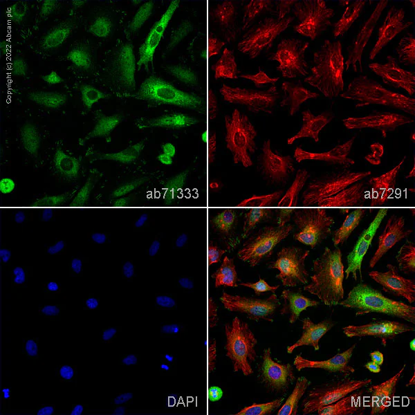
- ICC/IF
Lab
Immunocytochemistry/ Immunofluorescence - Anti-Talin 1 antibody (AB71333)
ab71333 staining Talin 1 in HeLa cells. The cells were fixed with 4% paraformaldehyde (10 min), permeabilized with 0.1% PBS-Tween for 5 minutes and then blocked with 1% BSA/10% normal goat serum/0.3M glycine in 0.1%PBS-Tween for 1h. The cells were then incubated overnight at 4°C with ab71333 at 1µg/ml and ab7291, Mouse monoclonal [DM1A] to alpha Tubulin - Loading Control. Cells were then incubated with ab150081, Goat polyclonal Secondary Antibody to Rabbit IgG - H&L (Alexa Fluor® 488), pre-adsorbed at 1/1000 dilution (shown in green) and ab150120, Goat polyclonal Secondary Antibody to Mouse IgG - H&L (Alexa Fluor® 594), pre-adsorbed at 1/1000 dilution (shown in pseudocolour red). Nuclear DNA was labelled with DAPI (shown in blue).
Image was acquired with a high-content analyser (Operetta CLS, Perkin Elmer) and a maximum intensity projection of confocal sections is shown.
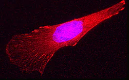
- ICC/IF
Collaborator
Immunocytochemistry/ Immunofluorescence - Anti-Talin 1 antibody (AB71333)
Image : Courtesy of Dr. Shaohua Li, UMDNJ-Robert Wood Johnson Medical School
Sample : mouse endoderm cell
Preparation :
Fix in 3% PFA in PBS for 30 min at RT
Primary antibody : Rabbit anti-talin (ab71333), 1 : 100
Secondary antibody : Goat polyclonal Secondary Antibody to Rabbit IgG - H&L (Cy3® 488) preadsorbed (ab97075), 1 : 100
Nuclear counterstain : DAPI
Rhodamine-phalloidin, 1 : 100
Nuclei were counterstained with DAPI
Courtesy of Dr. Shaohua Li, UMDNJ-Robert Wood Johnson Medical School

- WB
Lab
Western blot - Anti-Talin 1 antibody (AB71333)
Western blot : Anti-TLN1 antibody (ab71333) staining at 1 ug/ml, shown in green; Mouse anti-CANX [CANX/1543] (ab238078) loading control staining at 1/20000 dilution, shown in magenta. In Western blot, ab71333 was shown to bind specifically to TLN1. A band was observed at 270 kDa in wild-type PC-3 cell lysates with no signal observed at this size in TLN1 knockout cell line. To generate this image, wild-type and TLN1 knockout PC-3 cell lysates were analysed. First, samples were run on an SDS-PAGE gel then transferred onto a nitrocellulose membrane. Membranes were blocked in 3 % milk in TBS-0.1 % Tween® 20 (TBS-T) before incubation with primary antibodies overnight at 4 °C. Blots were washed four times in TBS-T, incubated with secondary antibodies for 1 h at room temperature, washed again four times then imaged. Secondary antibodies used were Goat anti-Rabbit IgG H&L 800CW and Goat anti-Mouse IgG H&L 680RD at 1/20000 dilution.
All lanes:
Western blot - Anti-Talin 1 antibody (ab71333) at 1 µg/mL
Lane 1:
Wild-type PC-3 cell lysate at 20 µg
Lane 2:
TLN1 knockout PC-3 cell lysate at 20 µg
Lane 3:
TLN1 knockout PC-3 cell lysate at 10 µg
Lane 4:
HeLa cell lysate at 20 µg
Lane 5:
SK-OV-3 cell lysate at 20 µg
Secondary
All lanes:
Goat anti-Rabbit IgG H&L 800CW and Goat anti-Mouse IgG H&L 680RD at 1/20000 dilution
Observed band size: 270 kDa
false
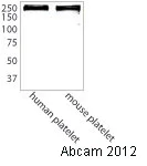
- WB
AbReview32678****
Western blot - Anti-Talin 1 antibody (AB71333)
Detection method : Licor System
All lanes:
Western blot - Anti-Talin 1 antibody (ab71333) at 1/1000 dilution
Lane 1:
Human platelets at 20 µg
Lane 2:
Mouse platelets at 20 µg
Secondary
All lanes:
Goat polyclonal to rabbit IgG conjugated to IRDye 800CW (undiluted)
Predicted band size: 269 kDa
false
Exposure time: 1min
This image is courtesy of an anonymous abreview.
- WB
Project
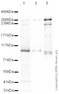
Western blot - Anti-Talin 1 antibody (AB71333)
All lanes:
Western blot - Anti-Talin 1 antibody (ab71333) at 1 µg/mL
Lane 1:
Jurkat (Human T cell lymphoblast-like cell line) Whole Cell Lysate at 10 µg
Lane 2:
Ramos (Human Burkitt's lymphoma cell line) Whole Cell Lysate at 10 µg
Lane 3:
MEF1 (Mouse embryonic fibroblast cell line) Whole Cell Lysate at 10 µg
Secondary
All lanes:
Goat polyclonal to Rabbit IgG - H&L - Pre-Adsorbed (HRP) at 1/3000 dilution
Predicted band size: 269 kDa
Observed band size: 100 kDa,230 kDa,270 kDa
false
Exposure time: 8min
替代产品
搭配使用产品
Primary Antibodies
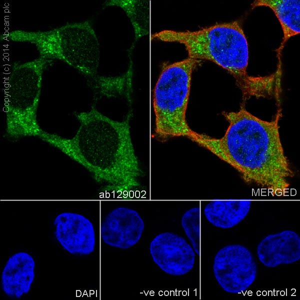
AB129002
Anti-Vinculin antibody [EPR8185] - Loading Control
20ul selling size|RabMAb|Recombinant|KO Validated
- 5
- 21
Primary Antibodies
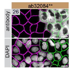
AB32084
BOND RX™ Validated|RabMAb|Recombinant|KO Validated|20ul selling size
- 5
- 21
Primary Antibodies
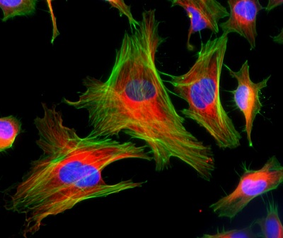
AB81298
Anti-FAK (phospho Y397) antibody [EP2160Y]
20ul selling size|RabMAb|Recombinant

- 4
- 2
反应性数据
性能和储存信息
形式
纯化工艺
存储溶液
运输条件
推荐的短期储存时间
推荐的短期储存条件
推荐的长期储存条件
分装信息
储存信息
补充信息
This supplementary information is collated from multiple sources and compiled automatically.
Biological function summary
Talin 1 is involved in facilitating integrin activation and focal adhesion formation. It is a component of a larger complex that includes proteins like vinculin and paxillin. These complexes are responsible for transmitting mechanical signals from the extracellular matrix into the intracellular environment initiating cellular responses like growth and differentiation. Talin 1 also modulates actin dynamics which contributes to changes in cell shape and motility. Its role in these processes highlights its significance in maintaining cellular architecture.
Pathways
Talin 1 participates in the integrin-mediated signaling pathway and the Rho GTPase signaling pathway. In the integrin pathway Talin 1 interacts with integrin beta subunits leading to integrin clustering and increased focal adhesion strength. In the Rho GTPase pathway Talin 1 influences actin cytoskeleton rearrangements through its interactions with proteins like RhoA and Rac1. These pathways are critical for processes such as cell proliferation and migration which are essential for proper tissue function and development.
产品实验方案
- Visit the General protocols
- Visit the Troubleshooting
靶点信息
文献 (44)
Recent publications for all applications. Explore the full list and refine your search
Science advances 11:eadt6083 PubMed40911671
2025
Applications
Unspecified application
Species
Unspecified reactive species
Nature 644:516-526 PubMed40681898
2025
Applications
Unspecified application
Species
Unspecified reactive species
Communications biology 8:948 PubMed40544224
2025
Applications
Unspecified application
Species
Unspecified reactive species
Discover oncology 16:374 PubMed40120027
2025
Applications
Unspecified application
Species
Unspecified reactive species
Heliyon 10:e36595 PubMed39286102
2024
Applications
Unspecified application
Species
Unspecified reactive species
PLoS computational biology 20:e1012341 PubMed39110765
2024
Applications
Unspecified application
Species
Unspecified reactive species
Science advances 9:eade2120 PubMed37585527
2023
Applications
Unspecified application
Species
Unspecified reactive species
eLife 12: PubMed37549051
2023
Applications
Unspecified application
Species
Unspecified reactive species
Iranian journal of pathology 18:312-326 PubMed37942198
2023
Applications
Unspecified application
Species
Unspecified reactive species
Tissue engineering. Part C, Methods 29:321-331 PubMed37416982
2023
Applications
Unspecified application
Species
Unspecified reactive species
Abcam Product Promise
Please note: All products are 'FOR RESEARCH USE ONLY. NOT FOR USE IN DIAGNOSTIC OR THERAPEUTIC PROCEDURES'.
For licensing inquiries, please contact partnerships@abcam.com