重组Anti-SNX3抗体[EPR28131-55]
Anti-SNX3 antibody [EPR28131-55]
- KO Validated
- KD Validated
- 20ul selling size
- RabMAb
- Recombinant
- 了解详情
Be the first to review this product! Submit a review
|
(0 Publication)
Rabbit Recombinant Monoclonal SNX3 antibody. Suitable for WB, ICC/IF, Flow Cyt (Intra) and reacts with Human, Mouse, Recombinant fragment - Human samples.
查看别名
Sorting nexin-3, Protein SDP3, SNX3

- Flow Cyt (Intra)
Supplier Data
Flow Cytometry (Intracellular) - Anti-SNX3 antibody [EPR28131-55] (AB314492)
Flow cytometric analysis of 4% paraformaldehyde fixed 90% methanol permeabilized THP-1 (human monocytic leukemia monocyte) cells labelling SNX3 with ab314492 at 1/500 dilution (0.1 ug)/Red (Red) compared with a Rabbit monoclonal IgG (ab172730) (Black) isotype control and an unlabelled control (cells without incubation with primary antibody and secondary antibody) (Blue). A Goat Anti-Rabbit IgG (Alexa Fluor® 488, ab150081) at 1/5000 dilution was used as the secondary antibody.

- ICC/IF
Supplier Data
Immunocytochemistry/ Immunofluorescence - Anti-SNX3 antibody [EPR28131-55] (AB314492)
Immunofluorescent analysis of 4% Paraformaldehyde-fixed, 0.1% TritonX-100 permeabilized THP-1 (human monocytic leukemia monocyte) cells labelling SNX3 with ab314492 at 1/100 (5.17 ug/ml) dilution, followed by ab150081 Goat Anti-Rabbit IgG H&L (Alexa Fluor® 488) preadsorbed antibody at 1/1000 2 ug/ml dilution (Green). Confocal image showing cytoplasmic staining in THP-1 cell line. Image was taken with a confocal microscope(Leica-Microsystems, TCS SP8). ab195889 Anti-alpha Tubulin mouse monoclonal antibody - Microtubule Marker (Alexa Fluor® 594) was used to counterstain tubulin at 1/200 2.5ug/ml dilution (Red). The Nuclear counterstain was DAPI (Blue). Secondary antibody only control : Secondary antibody is ab150081 Goat Anti-Rabbit IgG H&L (Alexa Fluor® 488) preadsorbed at 1/1000 2 ug/ml dilution.

- Flow Cyt (Intra)
Supplier Data
Flow Cytometry (Intracellular) - Anti-SNX3 antibody [EPR28131-55] (AB314492)
Flow cytometric analysis of 4% paraformaldehyde fixed 90% methanol permeabilized NIH/3T3 (mouse embryonic fibroblast) cells labelling SNX3 with ab314492 at 1/500 dilution (0.1 ug)/Red (Red) compared with a Rabbit monoclonal IgG (ab172730) (Black) isotype control and an unlabelled control (cells without incubation with primary antibody and secondary antibody) (Blue). A Goat Anti-Rabbit IgG (Alexa Fluor® 488, ab150081) at 1/5000 dilution was used as the secondary antibody.

- ICC/IF
Supplier Data
Immunocytochemistry/ Immunofluorescence - Anti-SNX3 antibody [EPR28131-55] (AB314492)
Immunofluorescent analysis of 4% Paraformaldehyde-fixed, 0.1% TritonX-100 permeabilized NIH/3T3 (mouse embryonic fibroblast) cells labelling SNX3 with ab314492 at 1/100 (5.17 ug/ml) dilution, followed by ab150081 Goat Anti-Rabbit IgG H&L (Alexa Fluor® 488) preadsorbed antibody at 1/1000 2 ug/ml dilution (Green). Confocal image showing cytoplasmic staining in NIH/3T3 cell line. Image was taken with a confocal microscope(Leica-Microsystems, TCS SP8). ab195889 Anti-alpha Tubulin mouse monoclonal antibody - Microtubule Marker (Alexa Fluor® 594) was used to counterstain tubulin at 1/200 2.5ug/ml dilution (Red). The Nuclear counterstain was DAPI (Blue). Secondary antibody only control : Secondary antibody is ab150081 Goat Anti-Rabbit IgG H&L (Alexa Fluor® 488) preadsorbed at 1/1000 2 ug/ml dilution.
- WB
Lab
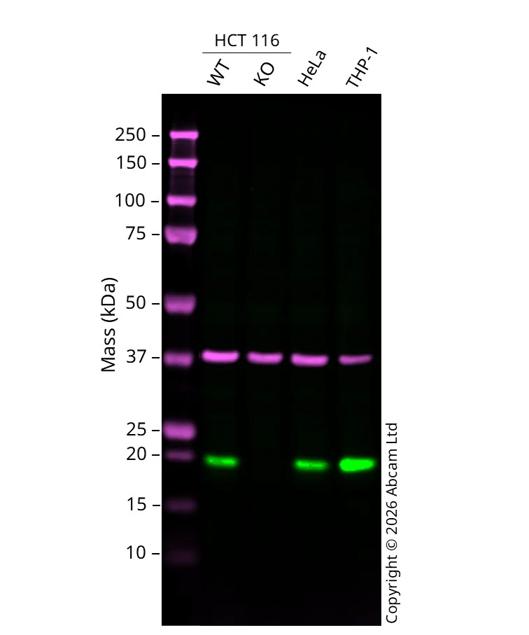
Western blot - Anti-SNX3 antibody [EPR28131-55] (AB314492)
Western blot : Anti-SNX3 antibody [EPR28131-55] ab314492 staining at 1/1000 dilution, shown in green; Mouse anti GAPDH ab8245 loading control staining at 1/20000 dilution, shown in magenta. A band was observed at 18 kDa in Wild-type HCT 116 cell lysates with no signal observed at this size in SNX3 knockout HCT 116 cell line. To generate this image, samples were run on an SDS-PAGE gel then transferred onto a nitrocellulose membrane. Membranes were blocked in 5% Milk in TBS-0.1 % Tween® 20 (TBS-T) before incubation with primary antibodies overnight at 4 °C. Blots were washed four times in TBS-T, incubated with secondary antibodies for 1 h at room temperature, washed again four times then imaged. Secondary antibodies used were Goat anti-Rabbit 800CW and Goat anti-Mouse 680RD at 1/20,000 dilution.
All lanes:
Western blot - Anti-SNX3 antibody [EPR28131-55] (ab314492) at 1/1000 dilution
Lane 1:
Western blot - Human wild-type HCT116 cell line (ab288559) at 20 µg
Lane 2:
SNX3 knockout HCT 116 at 20 µg
Lane 3:
HeLa at 20 µg
Lane 4:
THP-1 at 20 µg
Secondary
All lanes:
Goat anti-Rabbit 800CW & Goat anti-Mouse 680RD at 1/20000 dilution
Predicted band size: 18 kDa
Observed band size: 18 kDa
false

- WB
Supplier Data
Western blot - Anti-SNX3 antibody [EPR28131-55] (AB314492)
Blocking and diluting buffer and concentration : 5% NFDM/TBST
All lanes:
Western blot - Anti-SNX3 antibody [EPR28131-55] (ab314492) at 1/1000 dilution
Lane 1:
HeLa (human cervix adenocarcinoma epithelial cell) whole cell lysate at 20 µg
Lane 2:
NIH/3T3 (mouse embryonic fibroblast) whole cell lysate at 20 µg
Lane 3:
Neuro-2a (mouse neuroblastoma neuroblast) whole cell lysate at 20 µg
Secondary
All lanes:
Western blot - Goat Anti-Rabbit IgG H&L (HRP) (<a href='/products/secondary-antibodies/goat-rabbit-igg-h-l-hrp-ab97051'>ab97051</a>) at 1/100000 dilution
Observed band size: 18 kDa
false
Exposure time: 37s

- WB
Supplier Data
Western blot - Anti-SNX3 antibody [EPR28131-55] (AB314492)
Blocking and diluting buffer and concentration : 5% NFDM/TBST In Western blot, Anti-GAPDH antibody [EPR16891] - Loading Control (ab181602) staining at 1/200000 dilution.
All lanes:
Western blot - Anti-SNX3 antibody [EPR28131-55] (ab314492) at 1/1000 dilution
Lane 1:
THP-1 (human monocytic leukemia monocyte) transfected with scrambled siRNA control whole cell lysate at 20 µg
Lane 2:
THP-1 transfected with siRNA specifically targeti SNX3 whole cell lysate at 20 µg
Secondary
All lanes:
Western blot - Goat Anti-Rabbit IgG H&L (HRP) (<a href='/products/secondary-antibodies/goat-rabbit-igg-h-l-hrp-ab97051'>ab97051</a>) at 1/100000 dilution
Observed band size: 18 kDa
false
Exposure time: 26s

- WB
Supplier Data
Western blot - Anti-SNX3 antibody [EPR28131-55] (AB314492)
Blocking and diluting buffer and concentration : 5% NFDM/TBST.
This antibody does not cross-react with human SNX12.
This blot was developed using a high sensitivity ECL substrate. The high-sensitivity ECL substrate used allows for the detection of proteins in the mid-femtogram range.
In Western blot, Anti-6X His tag® antibody [EPR20547] - ChIP Grade (ab213204) staining at 1/5000 dilution
All lanes:
Western blot - Anti-SNX3 antibody [EPR28131-55] (ab314492) at 1/1000 dilution
Lane 1:
His-tagged human SNX3 recombinant protein at 200 ng
Lane 2:
His-tagged human SNX12 recombinant protein at 200 ng
Secondary
All lanes:
Western blot - Goat Anti-Rabbit IgG H&L (HRP) (<a href='/products/secondary-antibodies/goat-rabbit-igg-h-l-hrp-ab97051'>ab97051</a>) at 1/20000 dilution
Observed band size: 18 kDa
true
Exposure time: 180s
不同偶联物与剂型 (1)
-
Anti-SNX3 antibody [EPR28131-55] - BSA and Azide free
搭配使用产品


Primary Antibodies
AB183734
RabMAb|Recombinant|Lab Essentials|20ul selling size

- 5
- 24
Primary Antibodies
AB237703
20ul selling size|KO Validated|RabMAb|Recombinant

- 0
- 0
反应性数据
产品详情
Patented technology
Our RabMAb® technology is a patented hybridoma-based technology for making rabbit monoclonal antibodies. For details on our patents, please refer to RabMAb® patents.
What are the advantages of a recombinant monoclonal antibody?
This product is a recombinant monoclonal antibody, which offers several advantages including:
- - High batch-to-batch consistency and reproducibility
- - Improved sensitivity and specificity
- - Long-term security of supply
- - Animal-free batch production
For more information, read more on recombinant antibodies.
性能和储存信息
形式
纯化工艺
存储溶液
运输条件
推荐的短期储存时间
推荐的短期储存条件
推荐的长期储存条件
分装信息
储存信息
产品实验方案
- Visit the General protocols
- Visit the Troubleshooting
靶点信息
Abcam Product Promise
Please note: All products are 'FOR RESEARCH USE ONLY. NOT FOR USE IN DIAGNOSTIC OR THERAPEUTIC PROCEDURES'.
For licensing inquiries, please contact partnerships@abcam.com