重组Anti-Smad2 + Smad3抗体[18/Smad2/3]
Anti-Smad2 + Smad3 antibody [18/Smad2/3]
- BOND RX™ Validated
- Recombinant
- 了解详情
Be the first to review this product! Submit a review
|
(0 Publication)
Mouse Recombinant Monoclonal SMAD2 antibody. Suitable for IHC-P, WB and reacts with Rat, Mouse, Human samples.
查看别名
Madh2, Madr2, Smad2, SMAD family member 2, SMAD 2, Mad-related protein 2, Mothers against decapentaplegic homolog 2, mMad2, MAD homolog 2, Mothers against DPP homolog 2, Madh3, Smad3, SMAD family member 3, SMAD 3, Mothers against decapentaplegic homolog 3, MAD homolog 3, Mad3, Mothers against DPP homolog 3, mMad3
- IHC
Supplier Data
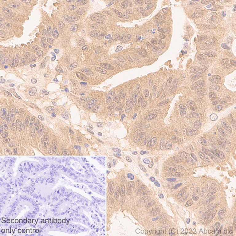
Immunohistochemistry - Anti-Smad2 + Smad3 antibody [18/Smad2/3] (AB305325)
Immunohistochemical analysis of paraffin-embedded human colon cancer tissue labeling Smad2 + Smad3 with ab305325 at 1/100 dilution (9.39 ug/ml) followed by ready to use LeicaDS9800 (Bond™ Polymer Refine Detection). Positive staining on human colon cancer is observed. The section was incubated with ab305325 for 30 mins at room temperature, followed by anti-mouse IgG1 antibody (ab125913) for 8 mins during the LeicaDS9800 kit staining procedure. The immunostaining was performed on a Leica Biosystems BOND® RX instrument. Counterstained with Hematoxylin. Secondary antibody only control : Secondary antibody is ready to use LeicaDS9800 (Bond™ Polymer Refine Detection). Heat mediated antigen retrieval was performed with Tris-EDTA buffer (pH 9.0, Epitope Retrieval Solution2) for 20 mins.
- IHC
Supplier Data
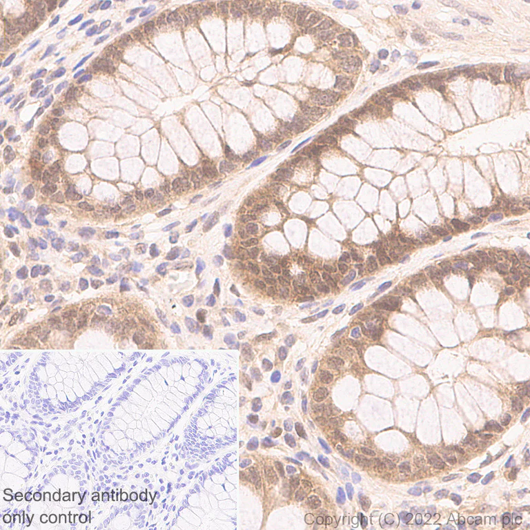
Immunohistochemistry - Anti-Smad2 + Smad3 antibody [18/Smad2/3] (AB305325)
Immunohistochemical analysis of paraffin-embedded human colon tissue labeling Smad2 + Smad3 with ab305325 at 1/100 dilution (9.39 ug/ml) followed by ready to use LeicaDS9800 (Bond™ Polymer Refine Detection). Positive staining on human colon is observed. The section was incubated with ab305325 for 30 mins at room temperature, followed by anti-mouse IgG1 antibody (ab125913) for 8 mins during the LeicaDS9800 kit staining procedure. The immunostaining was performed on a Leica Biosystems BOND® RX instrument. Counterstained with Hematoxylin. Secondary antibody only control : Secondary antibody is ready to use LeicaDS9800 (Bond™ Polymer Refine Detection). Heat mediated antigen retrieval was performed with Tris-EDTA buffer (pH 9.0, Epitope Retrieval Solution2) for 20 mins.
- IHC
Supplier Data
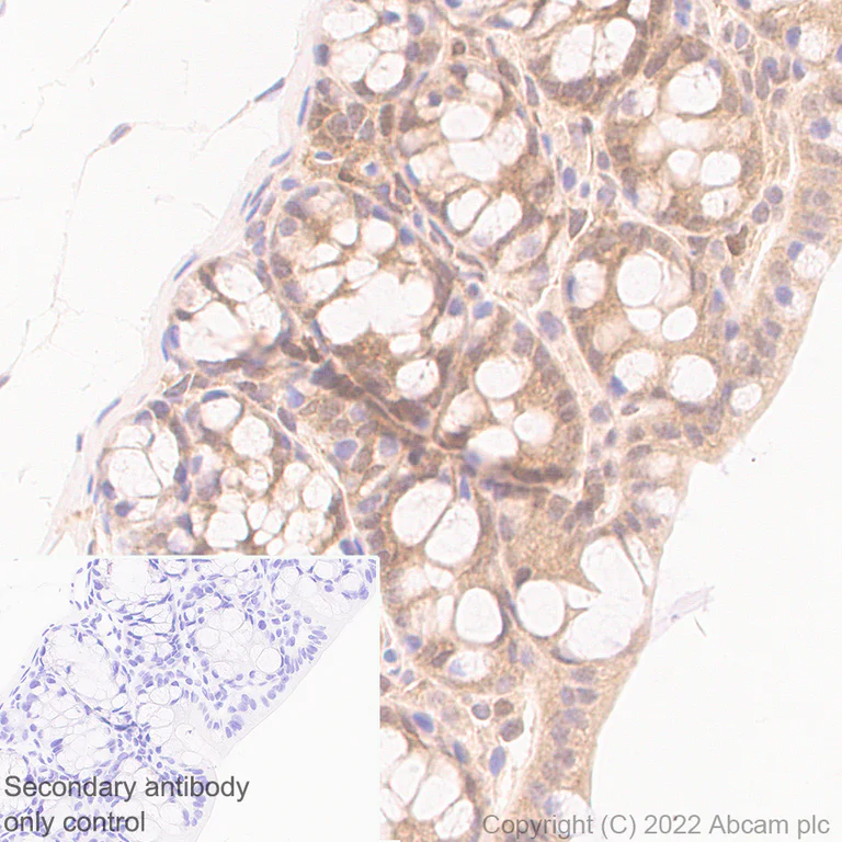
Immunohistochemistry - Anti-Smad2 + Smad3 antibody [18/Smad2/3] (AB305325)
Immunohistochemical analysis of paraffin-embedded mouse colon tissue labeling Smad2 + Smad3 with ab305325 at 1/250 dilution (3.756 ug/ml) followed by ready to use LeicaDS9800 (Bond™ Polymer Refine Detection). Positive staining on mouse colon is observed. The section was incubated with ab305325 for 30 mins at room temperature, followed by anti-mouse IgG1 antibody (ab125913) for 8 mins during the LeicaDS9800 kit staining procedure. The immunostaining was performed on a Leica Biosystems BOND® RX instrument. Counterstained with Hematoxylin. Secondary antibody only control : Secondary antibody is ready to use LeicaDS9800 (Bond™ Polymer Refine Detection). Heat mediated antigen retrieval was performed with Tris-EDTA buffer (pH 9.0, Epitope Retrieval Solution2) for 20 mins.
- IHC
Supplier Data
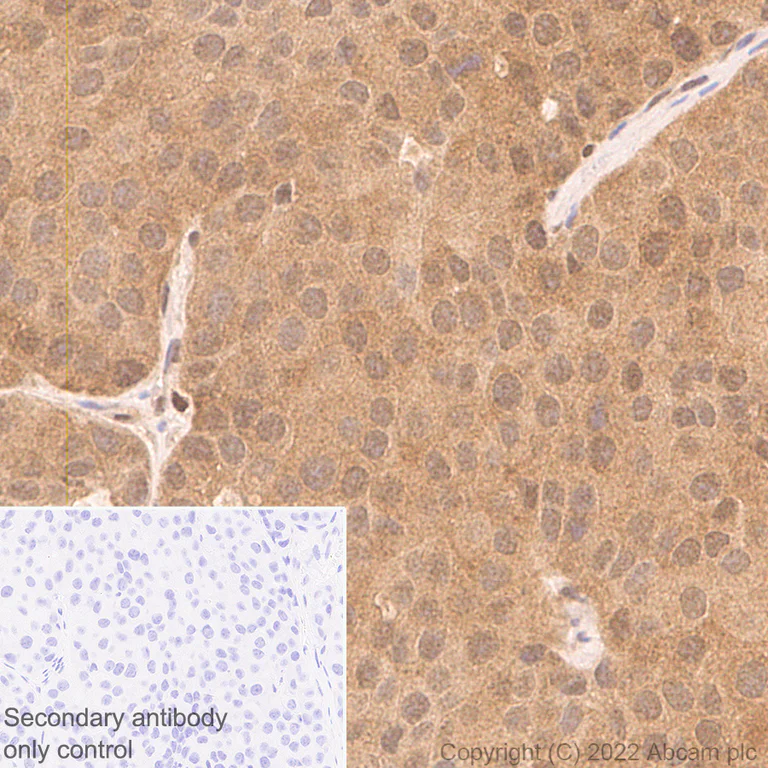
Immunohistochemistry - Anti-Smad2 + Smad3 antibody [18/Smad2/3] (AB305325)
Immunohistochemical analysis of paraffin-embedded mouse breast cancer tissue labeling Smad2 + Smad3 with ab305325 at 1/250 dilution (3.756 ug/ml) followed by ready to use LeicaDS9800 (Bond™ Polymer Refine Detection). Positive staining on mouse breast cancer is observed. The section was incubated with ab305325 for 30 mins at room temperature, followed by anti-mouse IgG1 antibody (ab125913) for 8 mins during the LeicaDS9800 kit staining procedure. The immunostaining was performed on a Leica Biosystems BOND® RX instrument. Counterstained with Hematoxylin. Secondary antibody only control : Secondary antibody is ready to use LeicaDS9800 (Bond™ Polymer Refine Detection). Heat mediated antigen retrieval was performed with Tris-EDTA buffer (pH 9.0, Epitope Retrieval Solution2) for 20 mins.
- IHC
Supplier Data
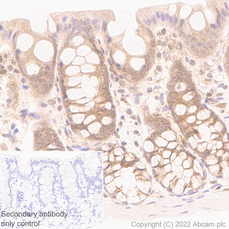
Immunohistochemistry - Anti-Smad2 + Smad3 antibody [18/Smad2/3] (AB305325)
Immunohistochemical analysis of paraffin-embedded rat colon tissue labeling Smad2 + Smad3 with ab305325 at 1/250 dilution (3.756 ug/ml) followed by ready to use LeicaDS9800 (Bond™ Polymer Refine Detection). Positive staining on rat colon is observed. The section was incubated with ab305325 for 30 mins at room temperature, followed by anti-mouse IgG1 antibody (ab125913) for 8 mins during the LeicaDS9800 kit staining procedure. The immunostaining was performed on a Leica Biosystems BOND® RX instrument. Counterstained with Hematoxylin. Secondary antibody only control : Secondary antibody is ready to use LeicaDS9800 (Bond™ Polymer Refine Detection). Heat mediated antigen retrieval was performed with Tris-EDTA buffer (pH 9.0, Epitope Retrieval Solution2) for 20 mins.
- WB
Supplier Data
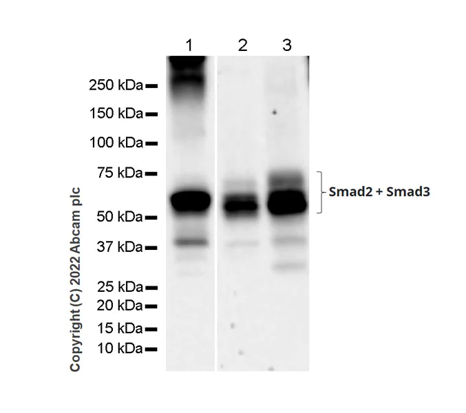
Western blot - Anti-Smad2 + Smad3 antibody [18/Smad2/3] (AB305325)
Blocking and diluting buffer and concentration : 5% NFDM/TBST. The expression profile/ molecular weight observed is consistent with what has been described in the literature (PMID : 18809571). The identity of the band around 37 kDa is unknown. Exposure times : Lanes 1, 4 and 5 : 180 seconds; Lanes 2 and 3 : 37 seconds.
All lanes:
Western blot - Anti-Smad2 + Smad3 antibody [18/Smad2/3] (ab305325) at 1/1000 dilution
Lane 1:
HeLa (human cervix adenocarcinoma epithelial cell), whole cell lysate 20 μg
Lane 2:
A549 (human lung carcinoma epithelial cell), whole cell lysate 20 μg
Lane 3:
HT-1080 (human fibrosarcoma epithelial cell), whole cell lysate 20 μg
Observed band size: 58 kDa,62 kDa
false
Exposure time: 114s
- WB
Supplier Data
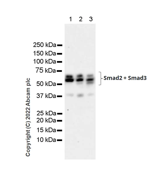
Western blot - Anti-Smad2 + Smad3 antibody [18/Smad2/3] (AB305325)
Blocking and diluting buffer and concentration : 5% NFDM/TBST. The expression profile/ molecular weight observed is consistent with what has been described in the literature (PMID : 18809571). The identity of the band around 37 kDa is unknown. Exposure time : 59 seconds.
All lanes:
Western blot - Anti-Smad2 + Smad3 antibody [18/Smad2/3] (ab305325) at 1/1000 dilution
Lane 1:
RAW264.7 (mouse Abelson murine leukemia virus-induced tumor macrophage), whole cell lysate 10 μg
Lane 2:
C2C12 (mouse myoblasts myoblast), whole cell lysate 10 μg
Lane 3:
C6 (rat glial tumor glial cell), whole cell lysate 10 μg
Observed band size: 58 kDa,62 kDa
false
Exposure time: 59s
- WB
Supplier Data
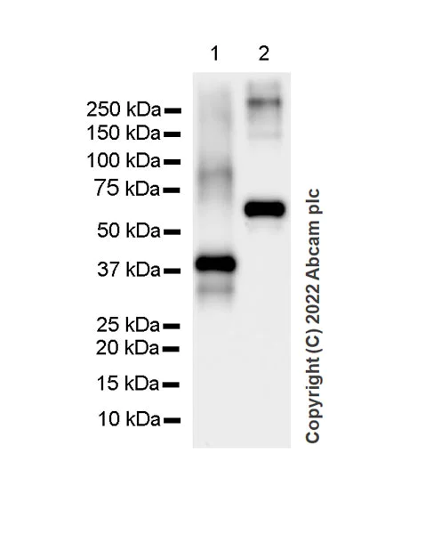
Western blot - Anti-Smad2 + Smad3 antibody [18/Smad2/3] (AB305325)
Blocking and diluting buffer and concentration : 5% NFDM/TBST. Exposure time : 15 seconds.
All lanes:
Western blot - Anti-Smad2 + Smad3 antibody [18/Smad2/3] (ab305325) at 1/1000 dilution
Lane 1:
His-tagged human Smad2 recombinant protein fragment 10ng
Lane 2:
His/GST-tagged human Smad3 recombinant protein fragment 10ng
Observed band size: 37 kDa,60 kDa
false
Exposure time: 15s
不同偶联物与剂型 (1)
-
Anti-Smad2 + Smad3 antibody [18/Smad2/3] - BSA and Azide free
搭配使用产品
Primary Antibodies
AB52903
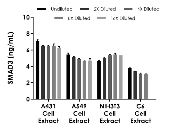
Anti-SMAD3 (pS423/425) + SMAD5 (pS463/465) + SMAD1 (pS463/465) + SMAD2 (pS465/467) antibody [EP823Y]
20ul selling size|Recombinant|Advanced Validation|RabMAb
- 4
- 21
Primary Antibodies
AB280888
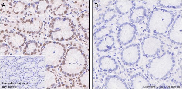
Anti-Smad2 (phospho S467) antibody [EPR23681-40]
BOND RX™ Validated|20ul selling size|RabMAb|Recombinant|KO Validated
- 5
- 2
Primary Antibodies
AB254407
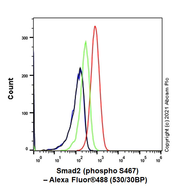
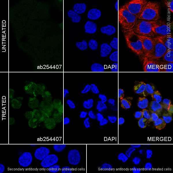
Anti-Smad2 (phospho T8) + Smad3 (phospho T8) antibody [EPR23682-64]
20ul selling size|Recombinant|Advanced Validation|RabMAb
- 0
- 0
Primary Antibodies
AB40854
20ul selling size|Recombinant|KO Validated|Advanced Validation|RabMAb

- 5
- 10
反应性数据
产品详情
Want a custom formulation?
This antibody clone is manufactured by Abcam. If you require a custom buffer formulation or conjugation for your experiments, please contact orders@abcam.com
What are the advantages of a recombinant monoclonal antibody?
This product is a recombinant monoclonal antibody, which offers several advantages including:
- - High batch-to-batch consistency and reproducibility
- - Improved sensitivity and specificity
- - Long-term security of supply
- - Animal-free batch production
For more information, read more on recombinant antibodies.
性能和储存信息
形式
纯化工艺
存储溶液
运输条件
推荐的短期储存时间
推荐的短期储存条件
推荐的长期储存条件
分装信息
储存信息
补充信息
This supplementary information is collated from multiple sources and compiled automatically.
Biological function summary
Smad2 and Smad3 regulate transcriptional responses essential for cellular processes including proliferation differentiation and apoptosis. These proteins form a trimeric complex with Smad4 upon activation by TGF-β receptors. The activated complex translocates into the nucleus where it binds to DNA and influences gene expression. Such actions highlight their fundamental role in controlling cell behavior and fate.
Pathways
Smad2 and Smad3 play critical roles within the TGF-β signaling pathway. This pathway has significant effects on cell cycle control and immune regulation. Both Smad2 and Smad3 interact closely with the protein Smad4 to mediate various downstream effects integrating signals with other pathways such as MAPK pathway which involves different cellular responses.
产品实验方案
- Visit the General protocols
- Visit the Troubleshooting
靶点信息
Abcam Product Promise
Please note: All products are 'FOR RESEARCH USE ONLY. NOT FOR USE IN DIAGNOSTIC OR THERAPEUTIC PROCEDURES'.
For licensing inquiries, please contact partnerships@abcam.com