重组Anti-S100 Calcium Binding蛋白A13/S100A13抗体[EPR4510]
Anti-S100 Calcium Binding Protein A13/S100A13 antibody [EPR4510]
- RabMAb
- Recombinant
- KO Validated
- 了解详情
Be the first to review this product! Submit a review
|
(5 Publications)
Rabbit Recombinant Monoclonal S100 Calcium Binding Protein A13/S100A13 antibody. Suitable for IHC-P, IP, WB, ICC/IF, Flow Cyt (Intra) and reacts with Human samples. Cited in 5 publications.
查看别名
Protein S100-A13, S100 calcium-binding protein A13, S100A13

- IHC-P
Unknown
Immunohistochemistry (Formalin/PFA-fixed paraffin-embedded sections) - Anti-S100 Calcium Binding Protein A13/S100A13 antibody [EPR4510] (AB109252)
ab109252, at a 1/250 dilution, staining S100 Calcium Binding Protein A13/S100A13 in Human brain, by Immunohistochemistry, Paraffin-embedded tissue.
Perform heat mediated antigen retrieval before commencing with IHC staining protocol.

- ICC/IF
Lab
Immunocytochemistry/ Immunofluorescence - Anti-S100 Calcium Binding Protein A13/S100A13 antibody [EPR4510] (AB109252)
Immunocytochemistry/ Immunofluorescence analysis of HUVEC (human umbilical vein endothelial cell) cells labeling S100 Calcium Binding Protein A13/S100A13 with purified ab109252 at 1/100 dilution (10 μg/mL). Cells were fixed in 4% Paraformaldehyde and permeabilized with 0.1% tritonX-100. Cells were counterstained with ab195889 Anti-alpha Tubulin antibody [DM1A] - Microtubule Marker (Alexa Fluor® 594) 1/200 (2.5 μg/mL). Goat anti rabbit IgG (Alexa Fluor® 488, ab150077) was used as the secondary antibody at 1/1000 (2 μg/mL) dilution. DAPI (blue) was used as nuclear counterstain. ab195889 Anti-alpha Tubulin antibody [DM1A] - Microtubule Marker (Alexa Fluor® 594) 1/200 (2.5 μg/mL) was used as the secondary antibody only control.

- Flow Cyt (Intra)
Lab
Flow Cytometry (Intracellular) - Anti-S100 Calcium Binding Protein A13/S100A13 antibody [EPR4510] (AB109252)
Intracellular Flow Cytometry analysis of HUVEC (Human umbilical vein endothelial cell) cells labeling S100 Calcium Binding Protein A13/S100A13 with purified ab109252 at 1/100 dilution (10 μg/mL) (Red). Cells were fixed with 4% Paraformaldehyde and permeabilised with 90% Methanol. A Goat anti rabbit IgG (Alexa Fluor® 488, ab150077) secondary antibody was used at 1/2000. Isotype control - Rabbit monoclonal IgG (Black). Unlabeled control - Cells without incubation with primary antibody and secondary antibody (Blue).

- IHC-P
Unknown
Immunohistochemistry (Formalin/PFA-fixed paraffin-embedded sections) - Anti-S100 Calcium Binding Protein A13/S100A13 antibody [EPR4510] (AB109252)
ab109252, at a 1/250 dilution, staining S100 Calcium Binding Protein A13/S100A13 in Human muscle, by Immunohistochemistry, Paraffin-embedded tissue.
Perform heat mediated antigen retrieval before commencing with IHC staining protocol.

- IP
Lab
Immunoprecipitation - Anti-S100 Calcium Binding Protein A13/S100A13 antibody [EPR4510] (AB109252)
Purified ab109252 at 1/50 dilution (2μg) immunoprecipitating S100 Calcium Binding Protein A13/S100A13 in Human heart lysate.
Lane 1 (input) : Human heart lysate 10μg
Lane 2 (+) : ab109252 + Human heart lysate.
Lane 3 (-) : Rabbit monoclonal IgG (ab172730) instead of ab109252 in Human heart lysate.
VeriBlot for IP Detection Reagent (HRP) (ab131366) (1/1000 dilution) was used for Western blotting.
Blocking Buffer and concentration : 5% NFDM/TBST.
Diluting buffer and concentration : 5% NFDM/TBST.
Observed band size : 11 kDa
All lanes:
Immunoprecipitation - Anti-S100 Calcium Binding Protein A13/S100A13 antibody [EPR4510] (ab109252)
Predicted band size: 11 kDa
false
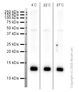
- WB
Lab
Western blot - Anti-S100 Calcium Binding Protein A13/S100A13 antibody [EPR4510] (AB109252)
This blot was produced using 14% SDS-PAGE containing 15 μg of HUVEC whole cell lysate per lane at 150V for 50 minutes before being transferred onto a 0.45 μm PVDF membrane at 72V for 50 minutes. The membrane was then blocked for 1hr using 5% NFDM/TBST, then incubated with ab109252 (1/1000) at room temperature for 1hr. After being washed three times in TBST, the membrane was incubated with Peroxidase conjugated goat anti-rabbit IgG at 1/2000 dilution for 1hr at room temperature. The membrane was washed three times again. Then the signal was developed using the ECL technique.
ab109252 was stored at a range of temperatures (+4°C, +22°C, +37°C) for 1 week before being tested in WB. The image shows the band intensity remains relatively constant across all storage temperatures, demonstrating that antibody activity is not affected under these conditions. This data was generated as part of a structured antibody stability study supporting Abcam’s transition from cold‑chain to ambient shipping.
All lanes:
Western blot - Anti-S100 Calcium Binding Protein A13/S100A13 antibody [EPR4510] (ab109252) at 1/1000 dilution
All lanes:
HUVEC whole cell lysate at 15 µg
Secondary
All lanes:
Western blot - Goat Anti-Rabbit IgG H&L (HRP) (<a href='/products/secondary-antibodies/goat-rabbit-igg-h-l-hrp-ab97051'>ab97051</a>) at 1/2000 dilution
false
Exposure time: 7s

- WB
Lab
Western blot - Anti-S100 Calcium Binding Protein A13/S100A13 antibody [EPR4510] (AB109252)
Lanes 1 - 4 : Merged signal (red and green). Green - ab109252 observed at 13 kDa. Red - loading control ab8245 (Mouse anti-GAPDH antibody [6C5]) observed at 37 kDa.
ab109252 was shown to react with S100 Calcium Binding Protein A13/S100A13 in wild-type HEK-293T cells in Western blot with loss of signal observed in S100A13 knockout cell line ab266065 (S100A13 knockout cell lysate ab258183). Wild-type HEK-293T and S100A13 knockout cell lysates were subjected to SDS-PAGE. Membranes were blocked in 3 % milk in TBS-T (0.1 % Tween®) before incubation with ab109252 and ab8245 (Mouse anti-GAPDH antibody [6C5]) overnight at 4°C at a 1 in 1000 dilution and a 1 in 20000 dilution respectively. Blots were incubated with Goat anti-Rabbit IgG H&L (IRDye® 800CW) preabsorbed (ab216773) and Goat anti-Mouse IgG H&L (IRDye® 680RD) preabsorbed (ab216776) secondary antibodies at 1 in 20000 dilution for 1 h at room temperature before imaging.
All lanes:
Western blot - Anti-S100 Calcium Binding Protein A13/S100A13 antibody [EPR4510] (ab109252) at 1/1000 dilution
Lane 1:
Wild-type HEK-293T cell lysate at 20 µg
Lane 2:
S100A13 knockout HEK-293T cell lysate at 20 µg
Lane 2:
Western blot - Human S100A13 (S100 Calcium Binding Protein A13) knockout HEK-293T cell line (<a href='/products/cell-lines/human-s100a13-s100-calcium-binding-protein-a13-knockout-hek-293t-cell-line-ab266065'>ab266065</a>)
Lane 3:
HeLa cell lysate at 20 µg
Lane 4:
Daudi cell lysate at 20 µg
Predicted band size: 11 kDa
Observed band size: 13 kDa
false

- WB
Unknown
Western blot - Anti-S100 Calcium Binding Protein A13/S100A13 antibody [EPR4510] (AB109252)
All lanes:
Western blot - Anti-S100 Calcium Binding Protein A13/S100A13 antibody [EPR4510] (ab109252) at 1/1000 dilution
Lane 1:
Human thyroid tissue lysate at 10 µg
Lane 2:
Human testis tissue lysate at 10 µg
Lane 3:
HUVEC lysate at 10 µg
Lane 4:
ECV304 lysate at 10 µg
Predicted band size: 11 kDa
false
不同偶联物与剂型 (1)
-
Anti-S100 Calcium Binding Protein A13/S100A13 antibody [EPR4510] - BSA and Azide free
替代产品

搭配使用产品
Primary Antibodies
AB52642
Anti-S100 beta antibody [EP1576Y] - Astrocyte Marker
BOND RX™ Validated|RabMAb|Recombinant|Lab Essentials|20ul selling size

- 4
- 37
Primary Antibodies
AB16667
BOND RX™ Validated|KO Validated|RabMAb|Advanced Validation|Recombinant|20ul selling size

- 5
- 133
Primary Antibodies
AB186735
Anti-TOMM20 antibody [EPR15581-54] - Mitochondrial Marker
RabMAb|Recombinant|Lab Essentials|20ul selling size

- 5
- 16
Primary Antibodies
AB177487
Anti-NeuN antibody [EPR12763] - Neuronal Marker
BOND RX™ Validated|RabMAb|Recombinant|Lab Essentials|20ul selling size|Advanced Validation

- 5
- 75
反应性数据
产品详情
Species reactivity
Mouse, Rat: We have preliminary internal testing data to indicate this antibody may not react with these species.
Please contact us for more information.
Patented technology
Our RabMAb® technology is a patented hybridoma-based technology for making rabbit monoclonal antibodies. For details on our patents, please refer to RabMAb® patents.
What are the advantages of a recombinant monoclonal antibody?
This product is a recombinant monoclonal antibody, which offers several advantages including:
- - High batch-to-batch consistency and reproducibility
- - Improved sensitivity and specificity
- - Long-term security of supply
- - Animal-free batch production
For more information, read more on recombinant antibodies.
性能和储存信息
形式
纯化工艺
存储溶液
运输条件
推荐的短期储存条件
推荐的长期储存条件
储存信息
补充信息
This supplementary information is collated from multiple sources and compiled automatically.
Biological function summary
S100A13 influences several cellular processes related to growth and differentiation. This protein facilitates secretion of fibroblast growth factor 1 (FGF1) and interleukin-1 alpha (IL-1α) through a non-classical pathway. S100A13 does not function alone; it engages in complexes with other proteins to execute its roles. It often works together with S100A6 and calcyclin-binding protein in these secretory pathways.
Pathways
S100A13 participates in the regulation of angiogenesis and inflammation. It is involved in the FGF signal transduction pathway by affecting the externalization of FGF1 an important player in angiogenesis. Additionally S100A13 contributes to inflammation-related processes. Its activity is linked to proteins such as p53 and FGF1 integrating it into wider cellular systems.
产品实验方案
- Visit the General protocols
- Visit the Troubleshooting
靶点信息
文献 (5)
Recent publications for all applications. Explore the full list and refine your search
Cell death & disease 16:563 PubMed40715046
2025
Applications
Unspecified application
Species
Unspecified reactive species
Cells 14: PubMed40497957
2025
Applications
Unspecified application
Species
Unspecified reactive species
Drug design, development and therapy 19:2609-2628 PubMed40196753
2025
Applications
Unspecified application
Species
Unspecified reactive species
Iranian journal of public health 51:2773-2782 PubMed36742226
2023
Applications
Unspecified application
Species
Unspecified reactive species
International journal of molecular sciences 23: PubMed35563653
2022
Applications
Unspecified application
Species
Unspecified reactive species
Abcam Product Promise
Please note: All products are 'FOR RESEARCH USE ONLY. NOT FOR USE IN DIAGNOSTIC OR THERAPEUTIC PROCEDURES'.
For licensing inquiries, please contact partnerships@abcam.com