Anti-Pax2 抗体 [EP3251]
Anti-Pax2 antibody [EP3251]
- 20ul selling size
- BOND RX™ Validated
- RabMAb
- Recombinant
- 了解详情
4
(11 Reviews)
|
(49 Publications)
Anti-Pax2 antibody [EP3251] (ab79389) is a rabbit monoclonal antibody detecting Pax2 in Western Blot, Flow Cytometry (Intra), IHC-P, ICC/IF. Suitable for Human, Mouse, Rat.
- Biophysical QC for unrivalled batch-batch consistency
- Over 30 publications
- Trusted since 2009
查看别名
Paired box protein Pax-2, PAX2
- IHC-P
Lab
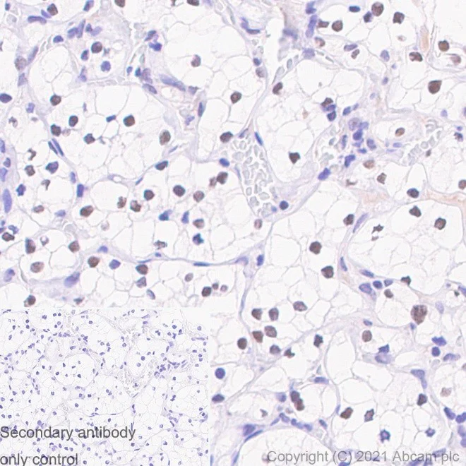
Immunohistochemistry (Formalin/PFA-fixed paraffin-embedded sections) - Anti-Pax2 antibody [EP3251] (AB79389)
Immunohistochemistry (Formalin/PFA-fixed paraffin-embedded sections) analysis of human renal cancer tissue labelling Pax2 with ab79389 at 1/100 dilution. Positive staining on human renal cancer. The section was incubated with ab79389 for 30 minutes at room temperature. Slides were incubated with 3% Hydrogen Peroxide for 10 mins at room temperature after secondary antibody incubation to reduce the background. The immunostaining was performed on a Leica Biosystems BOND® RX instrument. A Rabbit specific IHC polymer detection kit HRP/DAB (ab209101) was used as the secondary antibody (Ready-to-use). Counterstained with hematoxylin. Negative control using PBS instead of primary antibody.
Heat mediated antigen retrieval with Tris-EDTA buffer (pH 9.0, epitope retrieval solution 2) for 20 mins.

- Flow Cyt (Intra)
Supplier Data
Flow Cytometry (Intracellular) - Anti-Pax2 antibody [EP3251] (AB79389)
Intracellular Flow Cytometry analysis of K-562 (Human chronic myelogenous leukemia lymphoblast)labelling Pax2 with ab79389 at 1/30. Cells were fixed with 4% paraformaldehyde and permeabilised with 90% methanol. Goat Anti-Rabbit IgG H&L (Alexa Fluor® 488) (ab150077) at 1/2000 was used as the secondary antibody (red). Rabbit monoclonal IgG (ab172730) was used as the isotype control (black). Blue - Unlabelled control, cells without incubation with primary and secondary antibodies.

- IHC-P
Supplier Data
Immunohistochemistry (Formalin/PFA-fixed paraffin-embedded sections) - Anti-Pax2 antibody [EP3251] (AB79389)
Immunohistochemistry (Formalin/PFA-fixed paraffin-embedded sections) analysis of (A) human fetal kidney, (B) human normal kidney and (C) human renal cell carcinoma tissues labelled Pax2 with unpurified ab79389 at a dilution of 1/1000.

- IHC-P
Supplier Data
Immunohistochemistry (Formalin/PFA-fixed paraffin-embedded sections) - Anti-Pax2 antibody [EP3251] (AB79389)
Immunohistochemical analysis of Human malignant melanoma tissue, staining Pax2 with unpurified ab79389.
Antigen retrieval was performed via heat mediation in a citrate buffer (pH 6). Sections were blocked with 0.1% Triton X-100/PBS containing 1% BSA and 10% horse serum for 1 hour. Samples were incubated with primary antibody overnight at 4°C. A Cy3®-conjugated goat anti-rabbit IgG was used as the secondary antibody.

- IHC-P
Supplier Data
Immunohistochemistry (Formalin/PFA-fixed paraffin-embedded sections) - Anti-Pax2 antibody [EP3251] (AB79389)
Immunohistochemistry (Formalin/PFA-fixed paraffin-embedded sections) analysis of human kidney tissue labelling Pax2 with ab79389 at 1/100 dilution. Heat mediated antigen retrieval was performed using Tris/EDTA buffer pH 9. A Rabbit specific IHC polymer detection kit HRP/DAB (ab209101) was used as the secondary antibody (Ready-to-use). Negative control using PBS instead of primary antibody. Counterstained with hematoxylin. Positive staining on human kidney . The section was incubated with ab79389 for 30 mins at room temperature. The immunostaining was performed on a Leica Biosystems BOND® RX instrument

- ICC/IF
Supplier Data
Immunocytochemistry/ Immunofluorescence - Anti-Pax2 antibody [EP3251] (AB79389)
Immunocytochemistry analysis of HepG2 (human hepatocellular carcinoma epithelial cell) labelling Pax2 with ab79389 at 1/200 dilution. Cells were fixed with 4% paraformaldehyde and permeabilized with 0.1% Triton X-100 at room temperature for 4 minutes. Goat Anti-Rabbit IgG H&L (Alexa Fluor® 488) (ab150077) at 1/1000 was used as the secondary antibody (green). Cells were counterstained with Anti-alpha Tubulin antibody [DM1A] - Microtubule Marker (Alexa Fluor® 594) (ab195889) at 1/200 dilution (red). Nuclear DNA was labelled with DAPI (blue). Confocal image showing nucleolar staining in HepG2 cell line.
- IHC-P
PubMed
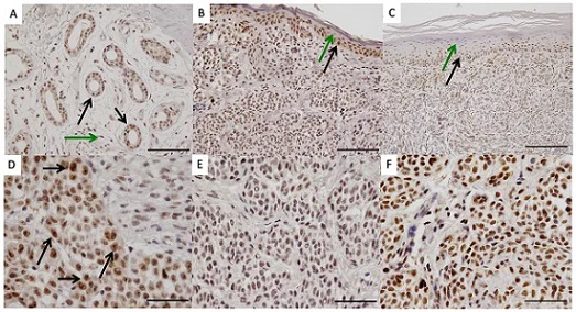
Immunohistochemistry (Formalin/PFA-fixed paraffin-embedded sections) - Anti-Pax2 antibody [EP3251] (AB79389)
Immunohistochemical analysis of Pax2 expression in tissue sections of human benign nevi and malignant melanoma using ab79389 at 1/100 dilution. All specimens were fixed in 4% formaline (pH 7.4), embedded in paraffin followed by cutting with a microtome (3 µm thickness). The slides were deparaffinized in xylol for 20 minutes and then rehydrated in descending series of ethanol (100%, 100%, 96%, 96%, 70%, and 70%). For antigen retrieval the slides were boiled in citrate buffer (pH 6.0) for 40 min, and then allowed to cool down for 15 min. After washing with PBS buffer the endogenous peroxidase was blocked with H2O2 for 15 min at room temperature. After washing in PBS the slides were incubated with the antibody against PAX2 (dilution 1/100) for 60 min at room temperature and washed in PBS again. The secondary antibody was incubated for 20 min at room temperature and after washing the slides in PBS the biotin streptavidine label was incubated for 20 min at room temperature. A detection kit including horseradish peroxidase and diaminobenzidine as chromogene was applied for 5 min. Counterstaining was performed with hematoxilin for 6 min. (A) In normal sweat glands, Pax2 is expressed in gland epithelial cells (black arrows) while intermingled stromal cells only show very weak or absent nuclear Pax2 expression (green arrow) Bar represents 100 µm. (B, C) Normal appearing epidermal cell layers adjacent to (B, bar represent 100 µm) nevi or (C, bar represent 200 µm) malignant melanoma show a differentially Pax2 expression with strongest Pax2 levels in germinal basal cell layers (black arrows) decreasing in higher differentiated keratinocytes and finally being absent in corneocytes (green arrows). (D) Malignant melanoma cells - heterogeneous nuclear Pax2 expression. Strongest expression in large atypical nuclei with prominent nucleoli (black arrows). (E, F) Pax2 expression in intradermal nevi was heterogeneous and did not correlate with histological features.
Lee SB et al. PAX2 regulates ADAM10 expression and mediates anchorage-independent cell growth of melanoma cells. PLoS One 6:e22312 (2011).
- ICC/IF
PubMed
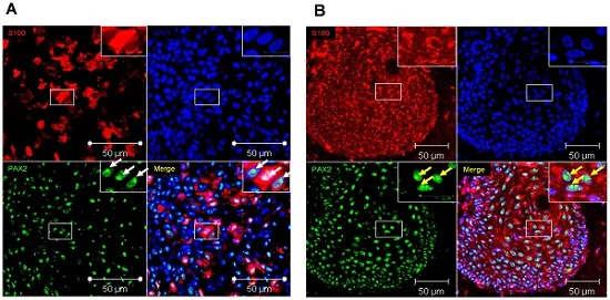
Immunocytochemistry/ Immunofluorescence - Anti-Pax2 antibody [EP3251] (AB79389)
Pax2 is expressed in melanocytes of benign nevi (A) and melanoma cells of patients with malignant melanoma (B). Cells were grown on coverslips and fixed with 4% paraformaldehyde/PBS. After washing the cells with PBS, cells were permeabilized and blocked with 0.1% Triton X-100/PBS containing 5% BSA. Pax2 (green) was examined by immunofluorescent analysis using ab79389 at 1/100 dilution (incubated for 1 hour at room temperature). Following 3 times washing, bound antibodies were deteced by Alexa 488 conjugated goat anti-mouse or Cy3 conjugated goat anti-mouse secondary antibodies. Following PBS-washing nuclei were stained with 4'6-diamidino-2-phenylindole (DAPI, Blue) and cells were mounted in Fluoromount-G™ and examined by fluorescence microscopy. White arrows in the higher magnified insets indicate PAX2 expression in nucleoli of melanocytes of benign nevi (A), yellow arrows in the higher magnified insets specify PAX2 expression in nucleoli of melanoma cells (B).
Lee SB et al. PAX2 regulates ADAM10 expression and mediates anchorage-independent cell growth of melanoma cells. PLoS One 6:e22312 (2011). Reproduced under the Creative Commons license http://creativecommons.org/licenses/by/4.0/

- IHC-P
Supplier Data
Immunohistochemistry (Formalin/PFA-fixed paraffin-embedded sections) - Anti-Pax2 antibody [EP3251] (AB79389)
Immunohistochemistry (Formalin/PFA-fixed paraffin-embedded sections) analysis of mouse kidney tissue labelling Pax2 with ab79389 at 1/100 dilution. Heat mediated antigen retrieval was performed using Tris/EDTA buffer pH 9. A Rabbit specific IHC polymer detection kit HRP/DAB (ab209101) was used as the secondary antibody (Ready-to-use). Negative control using PBS instead of primary antibody. Counterstained with hematoxylin. Positive staining on mouse kidney. The section was incubated with ab79389 for 30 mins at room temperature. The immunostaining was performed on a Leica Biosystems BOND® RX instrument.

- IHC-P
Supplier Data
Immunohistochemistry (Formalin/PFA-fixed paraffin-embedded sections) - Anti-Pax2 antibody [EP3251] (AB79389)
Immunohistochemistry (Formalin/PFA-fixed paraffin-embedded sections) analysis of rat kidney tissue labelling Pax2 with ab79389 at 1/100 dilution. Heat mediated antigen retrieval was performed using Tris/EDTA buffer pH 9. A Rabbit specific IHC polymer detection kit HRP/DAB (ab209101) was used as the secondary antibody (Ready-to-use). Negative control using PBS instead of primary antibody. Counterstained with hematoxylin. Positive staining on rat kidney tissue. The section was incubated with ab79389 for 30 mins at room temperature. The immunostaining was performed on a Leica Biosystems BOND® RX instrument.

- WB
Unknown
Western blot - Anti-Pax2 antibody [EP3251] (AB79389)
All lanes:
Western blot - Anti-Pax2 antibody [EP3251] (ab79389) at 1/10000 dilution
Lane 1:
HEK-293 (Human embryonic kidney epithelial cell) transfected with empty vector whole cell lysate at 15 µg
Lane 2:
HEK-293 (Human embryonic kidney epithelial cell) transfected with his tagged human full-length PAX2 at 15 µg
Secondary
All lanes:
Western blot - Goat Anti-Rabbit IgG H&L (HRP) (<a href='/products/secondary-antibodies/goat-rabbit-igg-h-l-hrp-ab97051'>ab97051</a>) at 1/20000 dilution
Predicted band size: 45 kDa
false
不同偶联物与剂型 (9)
-
Anti-Pax2 antibody [EP3251] - BSA and Azide free
-
660 APC
APC Anti-Pax2 antibody [EP3251]
-
519 Alexa Fluor® 488
Alexa Fluor® 488 Anti-Pax2 antibody [EP3251]
-
565 Alexa Fluor® 555
Alexa Fluor® 555 Anti-Pax2 antibody [EP3251]
-
603 Alexa Fluor® 568
Alexa Fluor® 568 Anti-Pax2 antibody [EP3251]
-
665 Alexa Fluor® 647
Alexa Fluor® 647 Anti-Pax2 antibody [EP3251]
-
775 Alexa Fluor® 750
Alexa Fluor® 750 Anti-Pax2 antibody [EP3251]
-
HRP Anti-Pax2 antibody [EP3251]
-
578 PE
PE Anti-Pax2 antibody [EP3251]
替代产品


Primary Antibodies
AB232460
Anti-Pax2 antibody [EPR8586] - BSA and Azide free
RabMAb|Recombinant

- 0
- 0
搭配使用产品
Primary Antibodies
AB89901
Anti-Wilms Tumor Protein antibody [CAN-R9(IHC)-56-2]
RabMAb|Recombinant|KO Validated|20ul selling size

- 5
- 43
Primary Antibodies
AB92547
Anti-Vimentin antibody [EPR3776] - Cytoskeleton Marker
BOND RX™ Validated|KO Validated|RabMAb|Recombinant|Lab Essentials|20ul selling size|Advanced Validation

- 5
- 92
Primary Antibodies
AB191870
Recombinant|Advanced Validation|RabMAb|20ul selling size

- 0
- 0
Primary Antibodies
AB195045
BOND RX™ Validated|RabMAb|Recombinant|20ul selling size

- 5
- 9
反应性数据
产品详情
What is this antibody validated in?
Anti-Pax2 antibody [EP3251] (ab79389) is a rabbit recombinant monoclonal antibody and is validated for use in Western Blot (WB), Flow Cytometry (Intra), Immunohistochemistry (IHC-P), Immunocytochemistry/immunofluorescence (ICC/IF) in Human, Mouse, Rat samples.
What is the molecular weight of Pax2?
Anti-Pax2 [EP3251] (ab79389) specifically detects a band for Pax2 (UniProt: Q02962) at a molecular weight of 45kDa.
Trusted by the scientific community
Anti-Pax2 [EP3251] (ab79389) was first used in a scientific publication in 2009 and has been cited over 30 times in peer-reviewed journals.
Reviewed by scientists
Anti-Pax2 [EP3251] (ab79389) has over 10 independent reviews from customers.
Other related products
We have a range of other formats of antibody clone [EP3251] also available for your convenience: ab79389, Alexa Fluor® 647 - ab218058, Carrier free - ab284763, PE - ab305674, APC - ab305675, HRP - ab305676, Alkaline Phosphatase - ab308810, Alexa Fluor® 488 - ab309759, Alexa Fluor® 555 - ab312072, Alexa Fluor® 568 - ab312549, Alexa Fluor® 750 - ab321734
Patented technology
Our RabMAb® technology is a patented hybridoma-based technology for making rabbit monoclonal antibodies. For details on our patents, please refer to RabMAb® patents.
What are the advantages of a recombinant monoclonal antibody?
This product is a recombinant monoclonal antibody, which offers several advantages including:
- - High batch-to-batch consistency and reproducibility
- - Improved sensitivity and specificity
- - Long-term security of supply
- - Animal-free batch production
For more information, read more on recombinant antibodies.
Trial sizes available!
Test your antibody or perform pre-screening before committing to a larger quantity. Sold in 20µl. Discover our selection of trial-size antibodies.
性能和储存信息
形式
纯化工艺
存储溶液
运输条件
推荐的短期储存时间
推荐的短期储存条件
推荐的长期储存条件
分装信息
储存信息
补充信息
This supplementary information is collated from multiple sources and compiled automatically.
Biological function summary
This transcription factor acts as a mediator in developmental processes influencing both organogenesis and tissue differentiation. Pax2 forms part of a transcriptional complex with other Pax family members such as Pax5 and Pax8 which together ensure the proper development of organs like the kidneys and eyes. These interactions allow Pax2 to control cell proliferation differentiation and survival in developing tissues thereby facilitating normal organ formation.
Pathways
Pax2 functions within the Wnt/Β-catenin and Sonic Hedgehog signaling pathways. These pathways are important for the regulation of cell fate and patterning during embryogenesis. Pax2 works closely with proteins such as Wnt1 and Gli1 to transduce signals that instruct the development of specific tissues. Pax2's involvement in these pathways demonstrates its critical role in responding to environmental cues that dictate developmental progression.
产品实验方案
- Visit the General protocols
- Visit the Troubleshooting
靶点信息
文献 (49)
Recent publications for all applications. Explore the full list and refine your search
Nature communications 16:8822 PubMed41044182
2025
Applications
Unspecified application
Species
Unspecified reactive species
Orphanet journal of rare diseases 20:289 PubMed40483479
2025
Applications
Unspecified application
Species
Unspecified reactive species
International journal of molecular sciences 25: PubMed39684657
2024
Applications
Unspecified application
Species
Unspecified reactive species
Investigative ophthalmology & visual science 65:34 PubMed39186263
2024
Applications
Unspecified application
Species
Unspecified reactive species
Stem cell research & therapy 15:4 PubMed38167128
2024
Applications
Unspecified application
Species
Unspecified reactive species
Nature communications 14:2378 PubMed37185772
2023
Applications
Unspecified application
Species
Unspecified reactive species
iScience 25:105555 PubMed36444301
2022
Applications
Unspecified application
Species
Unspecified reactive species
Molecular pain 18:17448069221108965 PubMed35815426
2022
Applications
Unspecified application
Species
Unspecified reactive species
Regenerative therapy 20:165-186 PubMed35620640
2022
Applications
Unspecified application
Species
Unspecified reactive species
Oncology letters 23:89 PubMed35126731
2022
Applications
Unspecified application
Species
Unspecified reactive species
Abcam Product Promise
Please note: All products are 'FOR RESEARCH USE ONLY. NOT FOR USE IN DIAGNOSTIC OR THERAPEUTIC PROCEDURES'.
For licensing inquiries, please contact partnerships@abcam.com