重组Anti-MBD3抗体[EPR18258]
Anti-MBD3 antibody [EPR18258]
- BOND RX™ Validated
- RabMAb
- Recombinant
- KO Validated
- 了解详情
Be the first to review this product! Submit a review
|
(2 Publications)
Rabbit Recombinant Monoclonal MBD3 antibody. Suitable for WB, ICC/IF, IHC-P and reacts with Mouse, Rat, Human samples. Cited in 2 publications.
查看别名
Methyl-CpG-binding domain protein 3, Methyl-CpG-binding protein MBD3, MBD3
- IHC-P
Supplier Data
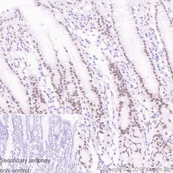
Immunohistochemistry (Formalin/PFA-fixed paraffin-embedded sections) - Anti-MBD3 antibody [EPR18258] (AB188401)
Immunohistochemical analysis of paraffin-embedded Human stomach tissue labeling MBD3 with ab188401 at 1/200 dilution (6.17 μg/ml) followed by a ready to use Rabbit specific IHC polymer detection kit HRP/DAB (ab209101). Nuclear staining on the human stomach. The section was incubated with ab188401 for 15 mins at room temperature. The immunostaining was performed on a Leica Biosystems BOND® RX instrument. Counterstained with Hematoxylin. Heat mediated antigen retrieval with Tris-EDTA buffer (pH 9.0 epitope retrieval solution 2) for 20 mins.
Secondary antibody only control : Used PBS instead of primary antibody secondary antibody is a ready to use Rabbit specific IHC polymer detection kit HRP/DAB (ab209101).
- ICC/IF
Lab
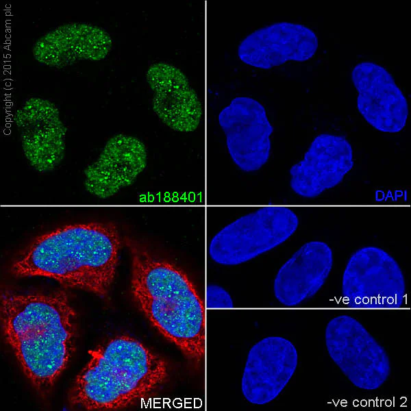
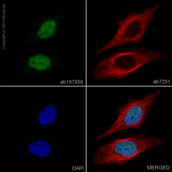
Immunocytochemistry/ Immunofluorescence - Anti-MBD3 antibody [EPR18258] (AB188401)
Immunofluorescent analysis of 4% paraformaldehyde-fixed 0.1% Triton X-100 permeabilized HeLa (Human epithelial cells from cervix adenocarcinoma) cells labeling MBD3 with ab188401 at 1/500 dilution followed by Goat anti-rabbit IgG (Alexa Fluor® 488) (ab150077) secondary antibody at 1/1000 dilution (green). Confocal image showing nuclear staining on HeLa cell line. The nuclear counter stain is DAPI (blue). Tubulin is detected with ab7291 (anti-Tubulin mouse mAb) at 1/1000 dilution and ab150120 (AlexaFluor®594 Goat anti-Mouse secondary) at 1/1000 dilution (red).
The negative controls are as follows :
-ve control 1 : ab188401 at 1/500 dilution followed by ab150120 (AlexaFluor®594 Goat anti-Mouse secondary) at 1/1000 dilution.
-ve control 2 : ab7291 (anti-Tubulin mouse mAb) at 1/1000 dilution followed by ab150077 (Alexa Fluor®488 Goat Anti-Rabbit IgG H&L) at 1/1000 dilution.
- IHC-P
Supplier Data
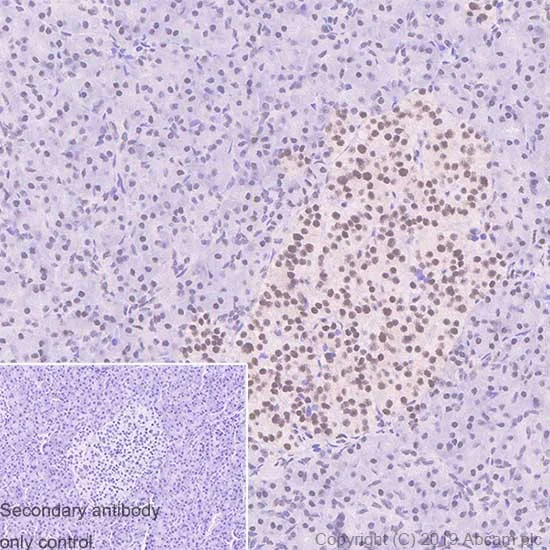
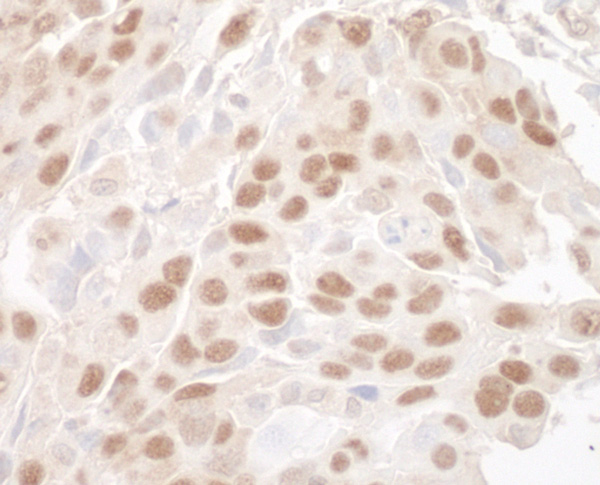
Immunohistochemistry (Formalin/PFA-fixed paraffin-embedded sections) - Anti-MBD3 antibody [EPR18258] (AB188401)
Immunohistochemical analysis of paraffin-embedded Human pancreas tissue labeling MBD3 with ab188401 at 1/200 dilution (6.17 μg/ml) followed by a ready to use Rabbit specific IHC polymer detection kit HRP/DAB (ab209101). Nuclear staining on the human pancreas (PMID : 27898661). The section was incubated with ab188401 for 15 mins at room temperature. The immunostaining was performed on a Leica Biosystems BOND® RX instrument. Counterstained with Hematoxylin. Heat mediated antigen retrieval with Tris-EDTA buffer (pH 9.0 epitope retrieval solution 2) for 20 mins.
Secondary antibody only control : Used PBS instead of primary antibody secondary antibody is a ready to use Rabbit specific IHC polymer detection kit HRP/DAB (ab209101).
- ICC/IF
Lab
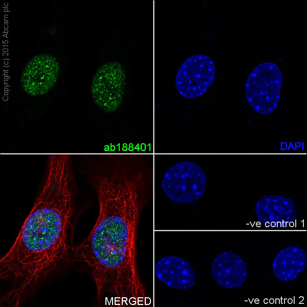
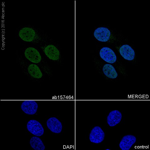
Immunocytochemistry/ Immunofluorescence - Anti-MBD3 antibody [EPR18258] (AB188401)
Immunofluorescent analysis of 4% paraformaldehyde-fixed 0.1% Triton X-100 permeabilized NIH/3T3 (Mouse embyro fibroblast cells) cells labeling MBD3 with ab188401 at 1/500 dilution followed by Goat anti-rabbit IgG (Alexa Fluor® 488) (ab150077) secondary antibody at 1/1000 dilution (green). Confocal image showing nuclear staining on NIH/3T3 cell line. The nuclear counter stain is DAPI (blue). Tubulin is detected with ab7291 (anti-Tubulin mouse mAb) at 1/1000 dilution and ab150120 (AlexaFluor®594 Goat anti-Mouse secondary) at 1/1000 dilution (red).
The negative controls are as follows :
-ve control 1 : ab188401 at 1/500 dilution followed by ab150120 (AlexaFluor®594 Goat anti-Mouse secondary) at 1/1000 dilution.
-ve control 2 : ab7291 (anti-Tubulin mouse mAb) at 1/1000 dilution followed by ab150077 (Alexa Fluor®488 Goat Anti-Rabbit IgG H&L) at 1/1000 dilution.
- WB
Lab
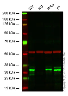
Western blot - Anti-MBD3 antibody [EPR18258] (AB188401)
Lane 1 : Wild-type HAP1 cell lysate (20 μg)
Lane 2 : MBD3 knockout HAP1 cell lysate (20 μg)
Lane 3 : HeLa cell lysate (20 μg)
Lane 4 : F9 cell lysate (20 μg)
Lanes 1 - 4 : Merged signal (red and green). Green - ab188401 observed at 33 kDa. Red - loading control, ab8245, observed at 37 kDa.
ab188401 was shown to specifically react with MBD3 when MBD3 knockout samples were used. Wild-type and MBD3 knockout samples were subjected to SDS-PAGE. ab188401 and ab8245 (loading control to GAPDH) were diluted 1/5000 and 1/10000 and incubated respectively overnight at 4°C. Blots were developed with Goat anti-Rabbit IgG H&L (IRDye® 800CW) preadsorbed (ab216773) and Goat anti-Mouse IgG H&L (IRDye® 680RD) preadsorbed (ab216776) secondary antibodies at 1/10000 dilution for 1 hour at room temperature before imaging.
All lanes:
Western blot - Anti-MBD3 antibody [EPR18258] (ab188401)
Predicted band size: 33 kDa
false
- WB
Supplier Data
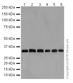
Western blot - Anti-MBD3 antibody [EPR18258] (AB188401)
Blocking/Dilution buffer : 5% NFDM/TBST.
The 33 and 29 kDa bands correspond to Isoforms 1 and 2.
All lanes:
Western blot - Anti-MBD3 antibody [EPR18258] (ab188401) at 1/5000 dilution
Lane 1:
HeLa (Human epithelial cells from cervix adenocarcinoma) whole cell lysate at 10 µg
Lane 2:
293 (Human epithelial cells from embryonic kidney) whole cell lysate at 10 µg
Lane 3:
F9 (Mouse embyro testicular cancer cell line) whole cell lysate at 10 µg
Lane 4:
WEHI-3 (Mouse leukemia cell line) whole cell lysate at 10 µg
Lane 5:
T-47D (Human ductal breast epithelial tumor cell line) whole cell lysate at 10 µg
Lane 6:
HepG2 (Human liver hepatocellular carcinoma) whole cell lysate at 10 µg
Secondary
All lanes:
Goat Anti-Rabbit IgG, (H+L), Peroxidase conjugated at 1/1000 dilution
Predicted band size: 33 kDa
Observed band size: 29 kDa,33 kDa
false
Exposure time: 1min
- WB
Supplier Data
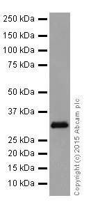
Western blot - Anti-MBD3 antibody [EPR18258] (AB188401)
Blocking/Dilution buffer : 5% NFDM/TBST.
All lanes:
Western blot - Anti-MBD3 antibody [EPR18258] (ab188401) at 1/2000 dilution
All lanes:
Human fetal kidney tissue lysate at 10 µg
Secondary
All lanes:
Anti-Rabbit IgG (HRP), specific to the non-reduced form of IgG at 1/1000 dilution
Predicted band size: 33 kDa
Observed band size: 29 kDa,33 kDa
false
Exposure time: 3min
- WB
Supplier Data
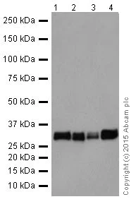
Western blot - Anti-MBD3 antibody [EPR18258] (AB188401)
Blocking/Dilution buffer : 5% NFDM/TBST.
The 33 and 29 kDa bands correspond to Isoforms 1 and 2.
All lanes:
Western blot - Anti-MBD3 antibody [EPR18258] (ab188401) at 1/2000 dilution
Lane 1:
C6 (Rat glial tumor cells) whole cell lysate at 10 µg
Lane 2:
RAW 264.7 (Mouse macrophage cells transformed with Abelson murine leukemia virus) whole cell lysate at 10 µg
Lane 3:
PC-12 (Rat adrenal gland pheochromocytoma) whole cell lysate at 10 µg
Lane 4:
NIH/3T3 (Mouse embyro fibroblast cells) whole cell lysate at 10 µg
Secondary
All lanes:
Goat Anti-Rabbit IgG, (H+L), Peroxidase conjugated at 1/1000 dilution
Predicted band size: 33 kDa
Observed band size: 29 kDa,33 kDa
false
Exposure time: 15s
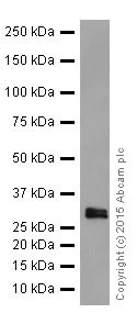
- WB
Supplier Data
Western blot - Anti-MBD3 antibody [EPR18258] (AB188401)
Blocking/Dilution buffer : 5% NFDM/TBST.
The 33 and 29 kDa bands correspond to Isoforms 1 and 2.
All lanes:
Western blot - Anti-MBD3 antibody [EPR18258] (ab188401) at 1/2000 dilution
All lanes:
Mouse spleen tissue lysate at 10 µg
Secondary
All lanes:
Goat Anti-Rabbit IgG, (H+L), Peroxidase conjugated at 1/1000 dilution
Predicted band size: 33 kDa
Observed band size: 29 kDa,33 kDa
false
Exposure time: 3min
不同偶联物与剂型 (1)
-
Anti-MBD3 antibody [EPR18258] - BSA and Azide free
替代产品
Primary Antibodies
AB157464
Anti-MBD3 antibody [EPR9913] - ChIP Grade
RabMAb|Recombinant|KO Validated
- 5
- 9
Primary Antibodies
AB240144
Anti-MBD3 antibody [EPR9913] - BSA and Azide free
RabMAb|Recombinant|KO Validated
- 0
- 0
搭配使用产品

Primary Antibodies
AB129195
Anti-KDM1/LSD1 antibody [EPR6825] - Nuclear Marker and ChIP Grade
KO Validated|RabMAb|Recombinant|Lab Essentials|20ul selling size|Advanced Validation

- 4
- 5
反应性数据
产品详情
Patented technology
Our RabMAb® technology is a patented hybridoma-based technology for making rabbit monoclonal antibodies. For details on our patents, please refer to RabMAb® patents.
What are the advantages of a recombinant monoclonal antibody?
This product is a recombinant monoclonal antibody, which offers several advantages including:
- - High batch-to-batch consistency and reproducibility
- - Improved sensitivity and specificity
- - Long-term security of supply
- - Animal-free batch production
For more information, read more on recombinant antibodies.
性能和储存信息
形式
纯化工艺
存储溶液
运输条件
推荐的短期储存时间
推荐的短期储存条件
推荐的长期储存条件
分装信息
储存信息
补充信息
This supplementary information is collated from multiple sources and compiled automatically.
Biological function summary
The activity of MBD3 impacts chromatin remodeling and gene expression regulation. MBD3 operates as a core component of the nucleosome remodeling deacetylase (NuRD) complex a multi-protein assembly important for transcriptional repression and chromatin structure modification. Its interaction within this complex helps modulate gene expression by altering the accessibility of chromatin and recruiting other modifiers for further regulation.
Pathways
MBD3 plays a significant role in key signaling and epigenetic pathways. It engages in pathways related to chromatin modification and transcription regulation. Its role in the NuRD complex underlines its participation in transcriptional regulation pathways impacting proteins such as HDAC1 and GATA2 which contribute to modifying histone acetylation and influencing gene transcription processes.
产品实验方案
- Visit the General protocols
- Visit the Troubleshooting
靶点信息
文献 (2)
Recent publications for all applications. Explore the full list and refine your search
The Journal of experimental medicine 217: PubMed32181805
2020
Applications
Unspecified application
Species
Unspecified reactive species
Immunity 50:121-136.e5 PubMed30594464
2018
Applications
Unspecified application
Species
Unspecified reactive species
Abcam Product Promise
Please note: All products are 'FOR RESEARCH USE ONLY. NOT FOR USE IN DIAGNOSTIC OR THERAPEUTIC PROCEDURES'.
For licensing inquiries, please contact partnerships@abcam.com