重组Anti-Glutaredoxin 1抗体[EPR29206-50] - BSA and Azide free
Anti-Glutaredoxin 1 antibody [EPR29206-50] - BSA and Azide free
- KD Validated
- BOND RX™ Validated
- Recombinant
- RabMAb
- 了解详情
Be the first to review this product! Submit a review
|
(0 Publication)
Rabbit Recombinant Monoclonal Glutaredoxin 1 antibody. Carrier free. Suitable for IHC-P, WB and reacts with Human, Mouse samples.
查看别名
GRX, GLRX, Glutaredoxin-1, Thioltransferase-1, TTase-1
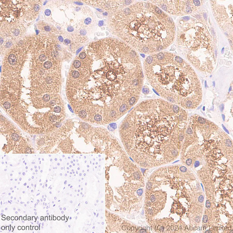
- IHC-P
Supplier Data
Immunohistochemistry (Formalin/PFA-fixed paraffin-embedded sections) - Anti-Glutaredoxin 1 antibody [EPR29206-50] - BSA and Azide free (AB319159)
This data was developed using ab319158, the same antibody clone in a different buffer formulation.
Immunohistochemical analysis of paraffin-embedded Human kidney tissue labeling Glutaredoxin 1 with ab319158 at 1/5000 (0.502 ug/ml) dilution, followed by a ready to use LeicaDS9800 (Bond™ Polymer Refine Detection).
Positive staining in human kidney (PMID : 11297543).
The section was incubated with ab319158 for 30 mins at room temperature.
The immunostaining was performed on a Leica Biosystems BOND® RX instrument
Counterstained with Hematoxylin.
MSD279 antibody only control : Secondary antibody is a ready to use LeicaDS9800 (Bond™ Polymer Refine Detection).
Heat mediated antigen retrieval was performed with Citrate buffer (pH 6.0, Epitope Retrieval Solution 1) for 20 mins
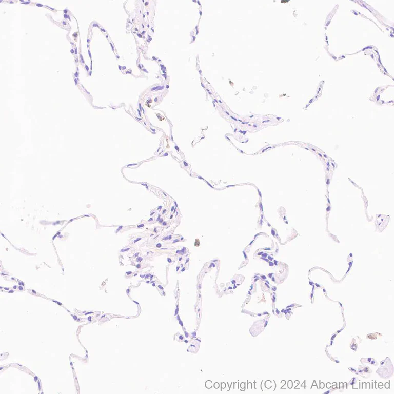
- IHC-P
Supplier Data
Immunohistochemistry (Formalin/PFA-fixed paraffin-embedded sections) - Anti-Glutaredoxin 1 antibody [EPR29206-50] - BSA and Azide free (AB319159)
This data was developed using ab319158, the same antibody clone in a different buffer formulation.
Immunohistochemical analysis of paraffin-embedded Human lung tissue labeling Glutaredoxin 1 with ab319158 at 1/5000 (0.1 ug/ml) dilution, followed by a ready to use LeicaDS9800 (Bond™ Polymer Refine Detection).
Low expression tissue : Nearly no staining in human lung.
The section was incubated with ab319158 for 30 mins at room temperature.
The immunostaining was performed on a Leica Biosystems BOND® RX instrument
Counterstained with Hematoxylin.
MSD279 antibody only control : Secondary antibody is a ready to use LeicaDS9800 (Bond™ Polymer Refine Detection).
Heat mediated antigen retrieval was performed with Tris-EDTA buffer (pH 9.0, Epitope Retrieval Solution2) for 20 mins
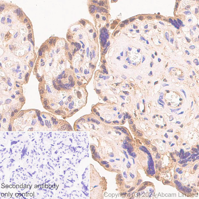
- IHC-P
Supplier Data
Immunohistochemistry (Formalin/PFA-fixed paraffin-embedded sections) - Anti-Glutaredoxin 1 antibody [EPR29206-50] - BSA and Azide free (AB319159)
This data was developed using ab319158, the same antibody clone in a different buffer formulation.
Immunohistochemical analysis of paraffin-embedded Human placenta tissue labeling Glutaredoxin 1 with ab319158 at 1/5000 (0.1 ug/ml) dilution, followed by a ready to use LeicaDS9800 (Bond™ Polymer Refine Detection).
Positive staining in human placenta (PMID : 11297543).
The section was incubated with ab319158 for 30 mins at room temperature.
The immunostaining was performed on a Leica Biosystems BOND® RX instrument
Counterstained with Hematoxylin.
MSD279 antibody only control : Secondary antibody is a ready to use LeicaDS9800 (Bond™ Polymer Refine Detection).
Heat mediated antigen retrieval was performed with Citrate buffer (pH 6.0, Epitope Retrieval Solution 1) for 20 mins
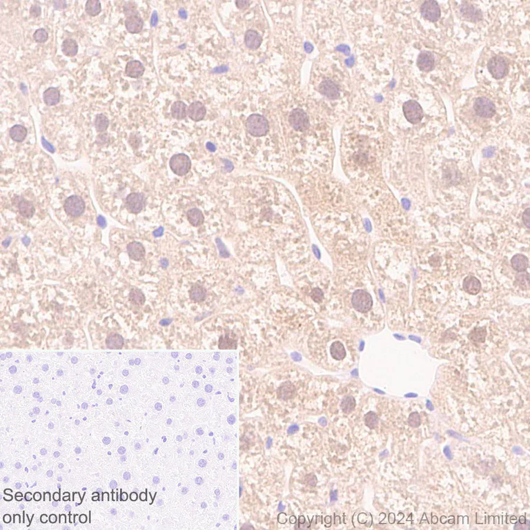
- IHC-P
Supplier Data
Immunohistochemistry (Formalin/PFA-fixed paraffin-embedded sections) - Anti-Glutaredoxin 1 antibody [EPR29206-50] - BSA and Azide free (AB319159)
This data was developed using ab319158, the same antibody clone in a different buffer formulation.
Immunohistochemical analysis of paraffin-embedded Mouse liver tissue labeling Glutaredoxin 1 with ab319158 at 1/1000 (0.502 ug/ml) dilution, followed by a ready to use LeicaDS9800 (Bond™ Polymer Refine Detection).
Positive staining in mouse liver.
The section was incubated with ab319158 for 30 mins at room temperature.
The immunostaining was performed on a Leica Biosystems BOND® RX instrument
Counterstained with Hematoxylin.
MSD279 antibody only control : Secondary antibody is a ready to use LeicaDS9800 (Bond™ Polymer Refine Detection).
Heat mediated antigen retrieval was performed with Citrate buffer (pH 6.0, Epitope Retrieval Solution 1) for 20 mins
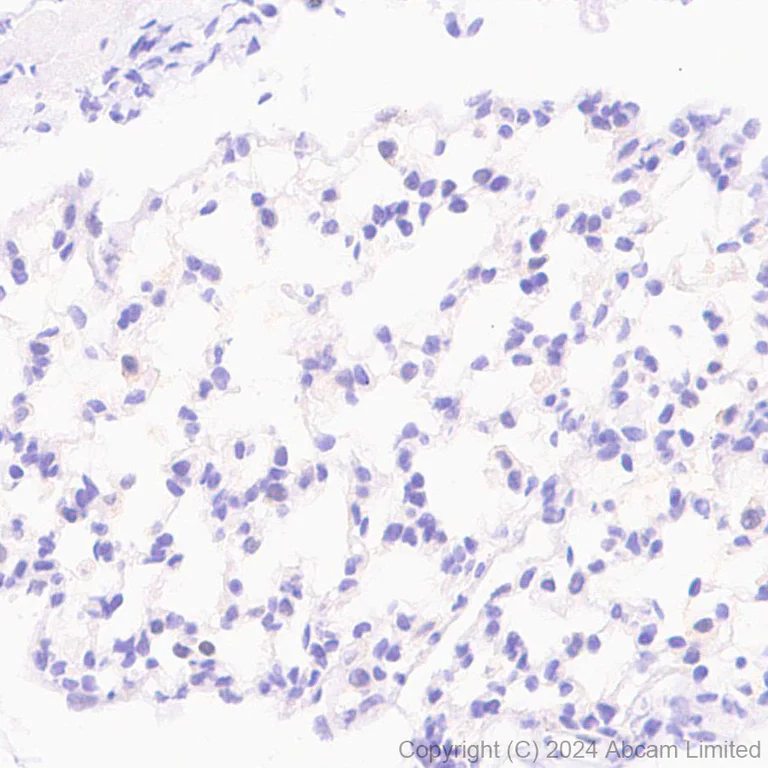
- IHC-P
Supplier Data
Immunohistochemistry (Formalin/PFA-fixed paraffin-embedded sections) - Anti-Glutaredoxin 1 antibody [EPR29206-50] - BSA and Azide free (AB319159)
This data was developed using ab319158, the same antibody clone in a different buffer formulation.
Immunohistochemical analysis of paraffin-embedded Mouse lung tissue labeling Glutaredoxin 1 with ab319158 at 1/1000 (0.502 ug/ml) dilution, followed by a ready to use LeicaDS9800 (Bond™ Polymer Refine Detection).
Low expression tissue : Nearly no staining in mouse lung.
The section was incubated with ab319158 for 30 mins at room temperature.
The immunostaining was performed on a Leica Biosystems BOND® RX instrument
Counterstained with Hematoxylin.
MSD279 antibody only control : Secondary antibody is a ready to use LeicaDS9800 (Bond™ Polymer Refine Detection).
Heat mediated antigen retrieval was performed with Tris-EDTA buffer (pH 9.0, Epitope Retrieval Solution2) for 20 mins
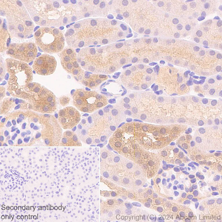
- IHC-P
Supplier Data
Immunohistochemistry (Formalin/PFA-fixed paraffin-embedded sections) - Anti-Glutaredoxin 1 antibody [EPR29206-50] - BSA and Azide free (AB319159)
This data was developed using ab319158, the same antibody clone in a different buffer formulation.
Immunohistochemical analysis of paraffin-embedded Mouse kidney tissue labeling Glutaredoxin 1 with ab319158 at 1/1000 (0.502 ug/ml) dilution, followed by a ready to use LeicaDS9800 (Bond™ Polymer Refine Detection).
Positive staining in mouse kidney.
The section was incubated with ab319158 for 30 mins at room temperature.
The immunostaining was performed on a Leica Biosystems BOND® RX instrument
Counterstained with Hematoxylin.
MSD279 antibody only control : Secondary antibody is a ready to use LeicaDS9800 (Bond™ Polymer Refine Detection).
Heat mediated antigen retrieval was performed with Citrate buffer (pH 6.0, Epitope Retrieval Solution 1) for 20 mins
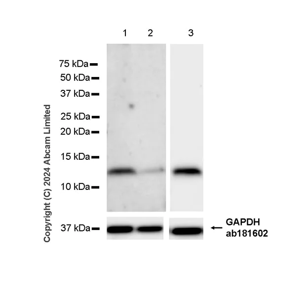
- WB
Supplier Data
Western blot - Anti-Glutaredoxin 1 antibody [EPR29206-50] - BSA and Azide free (AB319159)
This data was developed using ab319158, the same antibody clone in a different buffer formulation.
In Western blot, Anti-GAPDH antibody [EPR16891] - Loading Control (ab181602) staining at 1/200000 dilution.
Exposure time : Lanes 1-2 : 180 seconds, Lane 3 : 15 seconds
Blocking and dilution buffer and concentration : 5% NFDM/TBST
All lanes:
Western blot - Anti-Glutaredoxin 1 antibody [EPR29206-50] (<a href='/products/primary-antibodies/glutaredoxin-1-antibody-epr29206-50-ab319158'>ab319158</a>) at 1/1000 dilution
Lane 1:
HeLa (human cervical adenocarcinoma epithelial cell) transfected with scrambled siRNA control whole cell lysate at 20 µg
Lane 2:
Hela transfected with siRNA specifically targeting Glutaredoxin 1 whole cell lysate at 20 µg
Lane 3:
Jurkat (human T cell leukemia T lymphocyte from peripheral blood) whole cell lysate at 20 µg
Secondary
All lanes:
Goat Anti-Rabbit IgG (HRP) with minimal cross-reactivity with human IgG at 1/2000 dilution
Observed band size: 11 kDa,36 kDa
false
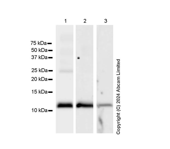
- WB
Supplier Data
Western blot - Anti-Glutaredoxin 1 antibody [EPR29206-50] - BSA and Azide free (AB319159)
This data was developed using ab319158, the same antibody clone in a different buffer formulation.
The identity of the higher MW band at approximately 25 kDa (in lane 1) is unknown.
Exposure time : Lanes 1 : 15 seconds, Lanes 2-3 : 92 seconds
Blocking and dilution buffer and concentration : 5% NFDM/TBST
All lanes:
Western blot - Anti-Glutaredoxin 1 antibody [EPR29206-50] (<a href='/products/primary-antibodies/glutaredoxin-1-antibody-epr29206-50-ab319158'>ab319158</a>) at 1/1000 dilution
Lane 1:
Human placenta tissue lysate at 20 µg
Lane 2:
Human skeletal muscle tissue lysate at 20 µg
Lane 3:
Mouse skeletal muscle tissue lysate at 20 µg
Secondary
All lanes:
Goat Anti-Rabbit IgG (HRP) with minimal cross-reactivity with human IgG at 1/2000 dilution
Observed band size: 11 kDa
false
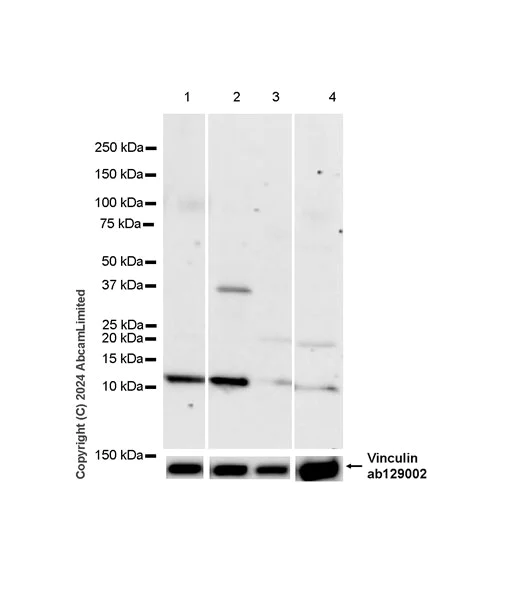
- WB
Supplier Data
Western blot - Anti-Glutaredoxin 1 antibody [EPR29206-50] - BSA and Azide free (AB319159)
This data was developed using ab319158, the same antibody clone in a different buffer formulation.
Low expression : brain, lung (PMID : 11297543).
The identity of the higher MW bands at approximately 37kDa (in lane 2) and 20kDa (in lanes 3-4) are unknown.
In Western blot, Anti-Vinculin antibody [EPR8185] (ab129002) staining at 1/10000 dilution.
Blocking and dilution buffer and concentration : 5% NFDM/TBST
All lanes:
Western blot - Anti-Glutaredoxin 1 antibody [EPR29206-50] (<a href='/products/primary-antibodies/glutaredoxin-1-antibody-epr29206-50-ab319158'>ab319158</a>) at 1/1000 dilution
Lane 1:
Mouse small intestine tissue lysate at 20 µg
Lane 2:
Mouse liver tissue lysate at 20 µg
Lane 3:
Mouse brain tissue lysate at 20 µg
Lane 4:
Mouse lung tissue lysate at 20 µg
Secondary
All lanes:
Western blot - Goat Anti-Rabbit IgG H&L (HRP) (<a href='/products/secondary-antibodies/goat-rabbit-igg-h-l-hrp-ab97051'>ab97051</a>) at 1/20000 dilution
Observed band size: 11 kDa,124 kDa
false
Exposure time: 92s
不同偶联物与剂型 (1)
-
Anti-Glutaredoxin 1 antibody [EPR29206-50]
搭配使用产品

Primary Antibodies
AB8227
Anti-beta Actin antibody - Loading Control
BOND RX™ Validated|Lab Essentials
- 5
- 104
反应性数据
产品详情
ab319159 is the carrier-free version of ab319158.
Patented technology
Our RabMAb® technology is a patented hybridoma-based technology for making rabbit monoclonal antibodies. For details on our patents, please refer to RabMAb® patents.
What are the advantages of a recombinant monoclonal antibody?
This product is a recombinant monoclonal antibody, which offers several advantages including:
- - High batch-to-batch consistency and reproducibility
- - Improved sensitivity and specificity
- - Long-term security of supply
- - Animal-free batch production
For more information, read more on recombinant antibodies.
Conjugation ready
Our carrier-free antibodies are typically supplied in a PBS-only formulation, purified and free of BSA, sodium azide and glycerol. This conjugation-ready format is designed for use with fluorochromes, metal isotopes, oligonucleotides, and enzymes, which makes them ideal for antibody labelling, functional and cell-based assays, flow-based assays (e.g. mass cytometry) and Multiplex Imaging applications.
Use our conjugation kits for antibody conjugates that are ready-to-use in as little as 20 minutes with 1 minute hands-on-time and 100% antibody recovery: available for fluorescent dyes, HRP, biotin and gold.
Compatibility
This product is compatible with the Maxpar® Antibody Labeling Kit from Fluidigm, without the need for antibody preparation. Maxpar® is a trademark of Fluidigm Canada Inc.
性能和储存信息
形式
纯化工艺
存储溶液
运输条件
推荐的短期储存条件
推荐的长期储存条件
补充信息
This supplementary information is collated from multiple sources and compiled automatically.
Biological function summary
This enzyme acts as an important regulator in cellular redox homeostasis and defense against oxidative stress. Glutaredoxin 1 does not technically form a complex but often interacts with multiple other proteins to modulate their redox state. It is involved in the regulation of apoptosis signaling pathways and the repair of oxidatively damaged proteins. Through these actions Glutaredoxin 1 protects cells from damage and maintains proper cellular function.
Pathways
Glutaredoxin 1 participates in key processes like the regulation of the thiol-disulfide balance and the glutathione metabolism pathway. These pathways are important for cellular detoxification and antioxidant defense. It works closely with glutathione peroxidase to mitigate oxidative damage and preserves cellular integrity by ensuring reduced forms of cysteine residues on proteins. This function links Glutaredoxin 1 to other proteins such as thioredoxin which shares similar roles in oxidative stress management.
产品实验方案
- Visit the General protocols
- Visit the Troubleshooting
靶点信息
Abcam Product Promise
Please note: All products are 'FOR RESEARCH USE ONLY. NOT FOR USE IN DIAGNOSTIC OR THERAPEUTIC PROCEDURES'.
For licensing inquiries, please contact partnerships@abcam.com