重组Anti-Factor H抗体[EPR26491-30]
Anti-Factor H antibody [EPR26491-30]
- 20ul selling size
- BOND RX™ Validated
- RabMAb
- Recombinant
- 了解详情
Be the first to review this product! Submit a review
|
(0 Publication)
Rabbit Recombinant Monoclonal Factor H antibody. Suitable for ICC/IF, WB, IHC-P and reacts with Mouse, Human, Rat samples.
查看别名
Hf1, Cfh, Complement factor H, Protein beta-1-H
- IHC-P
Supplier Data
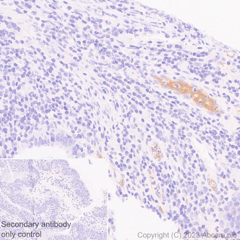
Immunohistochemistry (Formalin/PFA-fixed paraffin-embedded sections) - Anti-Factor H antibody [EPR26491-30] (AB315879)
Immunohistochemical analysis of paraffin-embedded Human colorectal carcinoma tissue labeling Factor H with ab315879 at 1/200 (0.976 ug/ml) dilution, followed by a ready to use LeicaDS9800 (Bond™ Polymer Refine Detection).
Positive staining on plasma of human colorectal carcinoma.
The section was incubated with ab315879 for 30 mins at room temperature.
The immunostaining was performed on a Leica Biosystems BOND® RX instrument
Counterstained with Hematoxylin.
Secondary antibody only control : Secondary antibody is a ready to use LeicaDS9800 (Bond™ Polymer Refine Detection).
Heat mediated antigen retrieval was performed with Tris-EDTA buffer (pH 9.0, Epitope Retrieval Solution2) for 20 mins
- IHC-P
Supplier Data
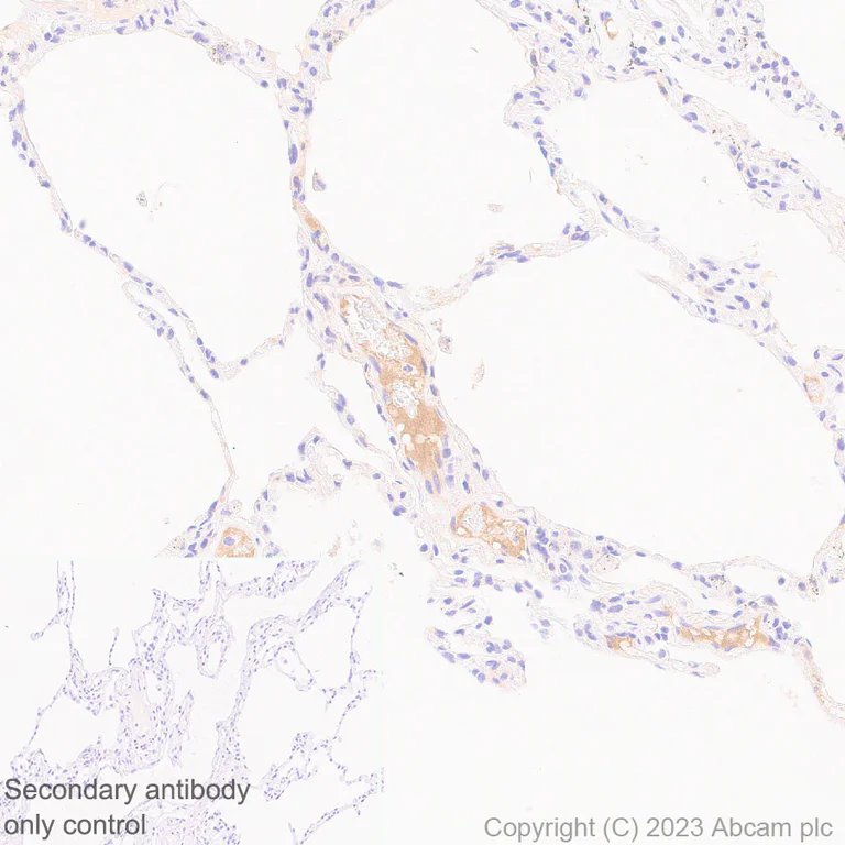
Immunohistochemistry (Formalin/PFA-fixed paraffin-embedded sections) - Anti-Factor H antibody [EPR26491-30] (AB315879)
Immunohistochemical analysis of paraffin-embedded Human lung tissue labeling Factor H with ab315879 at 1/200 (0.976 ug/ml) dilution, followed by a ready to use LeicaDS9800 (Bond™ Polymer Refine Detection).
Positive staining on plasma of human lung.
The section was incubated with ab315879 for 30 mins at room temperature.
The immunostaining was performed on a Leica Biosystems BOND® RX instrument
Counterstained with Hematoxylin.
Secondary antibody only control : Secondary antibody is a ready to use LeicaDS9800 (Bond™ Polymer Refine Detection).
Heat mediated antigen retrieval was performed with Tris-EDTA buffer (pH 9.0, Epitope Retrieval Solution2) for 20 mins
- IHC-P
Supplier Data
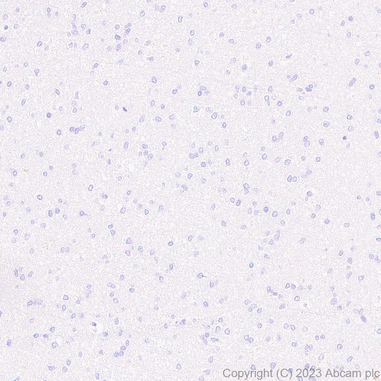
Immunohistochemistry (Formalin/PFA-fixed paraffin-embedded sections) - Anti-Factor H antibody [EPR26491-30] (AB315879)
Immunohistochemical analysis of paraffin-embedded Human cerebrum tissue labeling Factor H with ab315879 at 1/200 (0.976 ug/ml) dilution, followed by a ready to use LeicaDS9800 (Bond™ Polymer Refine Detection).
Negative control : no staining on human cerebrum.
The section was incubated with ab315879 for 30 mins at room temperature.
The immunostaining was performed on a Leica Biosystems BOND® RX instrument
Counterstained with Hematoxylin.
Secondary antibody only control : Secondary antibody is a ready to use LeicaDS9800 (Bond™ Polymer Refine Detection).
Heat mediated antigen retrieval was performed with Tris-EDTA buffer (pH 9.0, Epitope Retrieval Solution2) for 20 mins
- IHC-P
Supplier Data
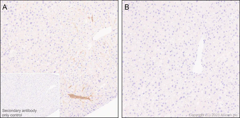
Immunohistochemistry (Formalin/PFA-fixed paraffin-embedded sections) - Anti-Factor H antibody [EPR26491-30] (AB315879)
Immunohistochemical analysis of paraffin-embedded (A) Mouse non-perfused fixed liver (B) Mouse perfused fixed liver tissue labeling Factor H with ab315879 at 1/500 (0.976 ug/ml) dilution, followed by a ready to use LeicaDS9800 (Bond™ Polymer Refine Detection).
Positive staining on (A) plasma of non-perfused fixed mouse liver. No staining on (B) perfused fixed mouse liver. (PMID : 32369457).
The section was incubated with ab315879 for 30 mins at room temperature.
The immunostaining was performed on a Leica Biosystems BOND® RX instrument
Counterstained with Hematoxylin.
Secondary antibody only control : Secondary antibody is a ready to use LeicaDS9800 (Bond™ Polymer Refine Detection).
Heat mediated antigen retrieval was performed with Tris-EDTA buffer (pH 9.0, Epitope Retrieval Solution2) for 20 mins
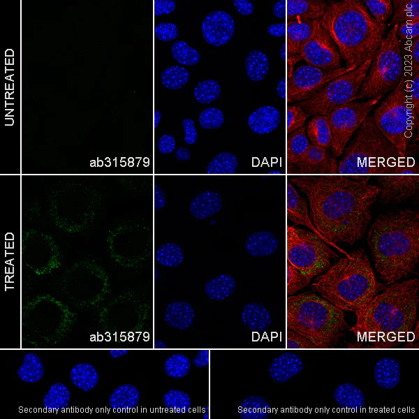
- ICC/IF
Supplier Data
Immunocytochemistry/ Immunofluorescence - Anti-Factor H antibody [EPR26491-30] (AB315879)
Immunofluorescent analysis of 4% Paraformaldehyde-fixed, 0.1% TritonX-100 permeabilized L-929 (mouse connective tissue fibroblast) cells labelling Factor H with ab315879 at 1/50 (9.76 ug/ml) dilution, followed by ab150081 Goat Anti-Rabbit IgG H&L (Alexa Fluor® 488) preadsorbed antibody at 1/1000 2 ug/ml dilution (Green).
Confocal image showing cytoplasmic staining in L-929 cells treated with IFN-gamma (500 units/ml) and dexamethasone (10 μM) for 24 hr. Image was taken with a confocal microscope(Leica-Microsystems, TCS SP8).
ab195889 Anti-alpha Tubulin mouse monoclonal antibody - Microtubule Marker (Alexa Fluor® 594) was used to counterstain tubulin at 1/200 2.5ug/ml dilution (Red). The Nuclear counterstain was DAPI (Blue).
Secondary antibody only control : Secondary antibody is ab150081 Goat Anti-Rabbit IgG H&L (Alexa Fluor® 488) preadsorbed at 1/1000 2 ug/ml dilution.
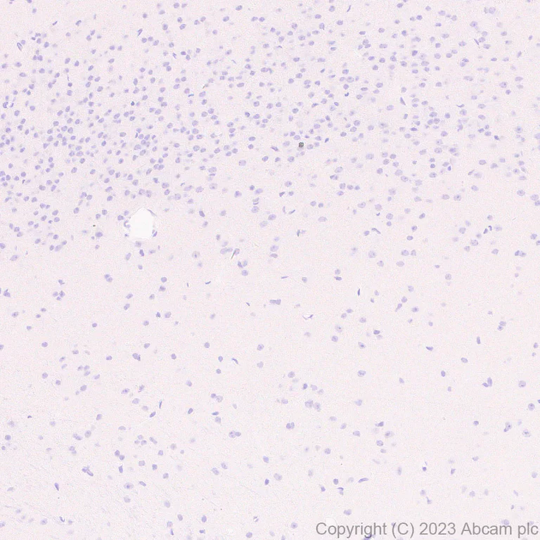
- IHC-P
Supplier Data
Immunohistochemistry (Formalin/PFA-fixed paraffin-embedded sections) - Anti-Factor H antibody [EPR26491-30] (AB315879)
Immunohistochemical analysis of paraffin-embedded Mouse cerebrum tissue labeling Factor H with ab315879 at 1/500 (0.976 ug/ml) dilution, followed by a ready to use LeicaDS9800 (Bond™ Polymer Refine Detection).
Negative control : no staining on mouse cerebrum.
The section was incubated with ab315879 for 30 mins at room temperature.
The immunostaining was performed on a Leica Biosystems BOND® RX instrument
Counterstained with Hematoxylin.
Secondary antibody only control : Secondary antibody is a ready to use LeicaDS9800 (Bond™ Polymer Refine Detection).
Heat mediated antigen retrieval was performed with Tris-EDTA buffer (pH 9.0, Epitope Retrieval Solution2) for 20 mins
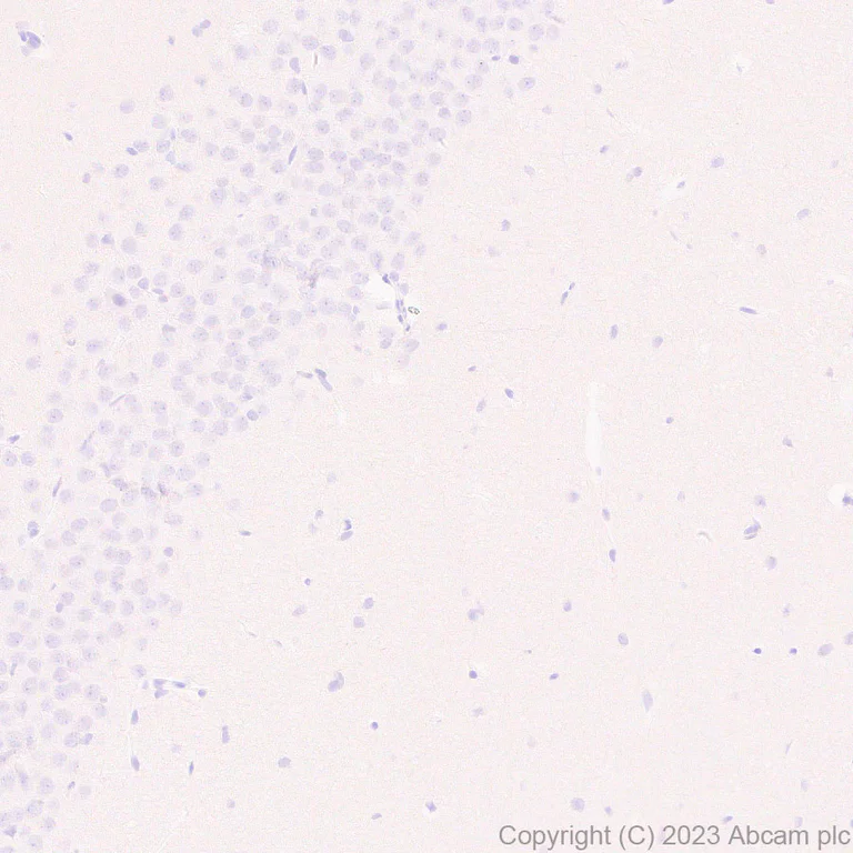
- IHC-P
Supplier Data
Immunohistochemistry (Formalin/PFA-fixed paraffin-embedded sections) - Anti-Factor H antibody [EPR26491-30] (AB315879)
Immunohistochemical analysis of paraffin-embedded Rat cerebrum tissue labeling Factor H with ab315879 at 1/500 (0.976 ug/ml) dilution, followed by a ready to use LeicaDS9800 (Bond™ Polymer Refine Detection).
Negative control : no staining on rat cerebrum.
The section was incubated with ab315879 for 30 mins at room temperature.
The immunostaining was performed on a Leica Biosystems BOND® RX instrument
Counterstained with Hematoxylin.
Secondary antibody only control : Secondary antibody is a ready to use LeicaDS9800 (Bond™ Polymer Refine Detection).
Heat mediated antigen retrieval was performed with Tris-EDTA buffer (pH 9.0, Epitope Retrieval Solution2) for 20 mins

- IHC-P
Supplier Data
Immunohistochemistry (Formalin/PFA-fixed paraffin-embedded sections) - Anti-Factor H antibody [EPR26491-30] (AB315879)
Immunohistochemical analysis of paraffin-embedded (A) Rat non-perfused fixed liver (B) Rat perfused fixed liver tissue labeling Factor H with ab315879 at 1/500 (0.976 ug/ml) dilution, followed by a ready to use LeicaDS9800 (Bond™ Polymer Refine Detection).
Positive staining on (A) plasma of non-perfused fixed rat liver. No staining on (B) perfused fixed rat liver.
The section was incubated with ab315879 for 30 mins at room temperature.
The immunostaining was performed on a Leica Biosystems BOND® RX instrument
Counterstained with Hematoxylin.
Secondary antibody only control : Secondary antibody is a ready to use LeicaDS9800 (Bond™ Polymer Refine Detection).
Heat mediated antigen retrieval was performed with Tris-EDTA buffer (pH 9.0, Epitope Retrieval Solution2) for 20 mins
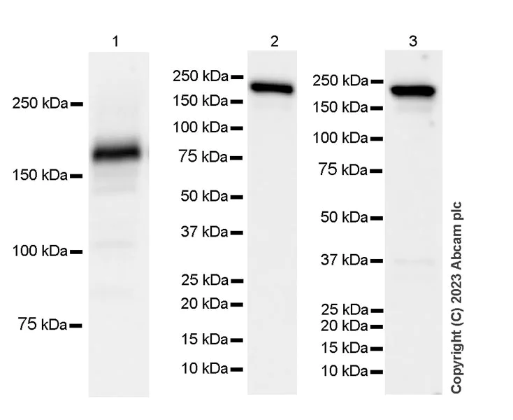
- WB
Supplier Data
Western blot - Anti-Factor H antibody [EPR26491-30] (AB315879)
Blocking and diluting buffer and concentration : 5% NFDM/TBST.
Samples are non-boiled as boiling may cause protein aggregation.
Lanes 1 and 3 are applied with Goat Anti-Rabbit IgG, (H+L), Peroxidase conjugated (ab97051) at 1/100000 and lane 2 is applied with Goat Anti-Rabbit IgG (HRP) with minimal cross-reactivity with human IgG at 1/2000.
Exposure time : Lane 1 : 6 seconds, lanes 2-3 : 15 seconds.
All lanes:
Western blot - Anti-Factor H antibody [EPR26491-30] (ab315879) at 1/1000 dilution
Lane 1:
Rat plasma lysate at 20 µg
Lane 2:
Human plasma lysate at 20 µg
Lane 3:
Mouse plasma lysate at 20 µg
Secondary
Lanes 1 and 3:
Western blot - Goat Anti-Rabbit IgG H&L (HRP) (<a href='/products/secondary-antibodies/goat-rabbit-igg-h-l-hrp-ab97051'>ab97051</a>) at 1/100000 dilution
Lane 2:
Goat Anti-Rabbit IgG (HRP) with minimal cross-reactivity with human IgG at 1/2000 dilution
Observed band size: 160 kDa
false
- WB
Supplier Data
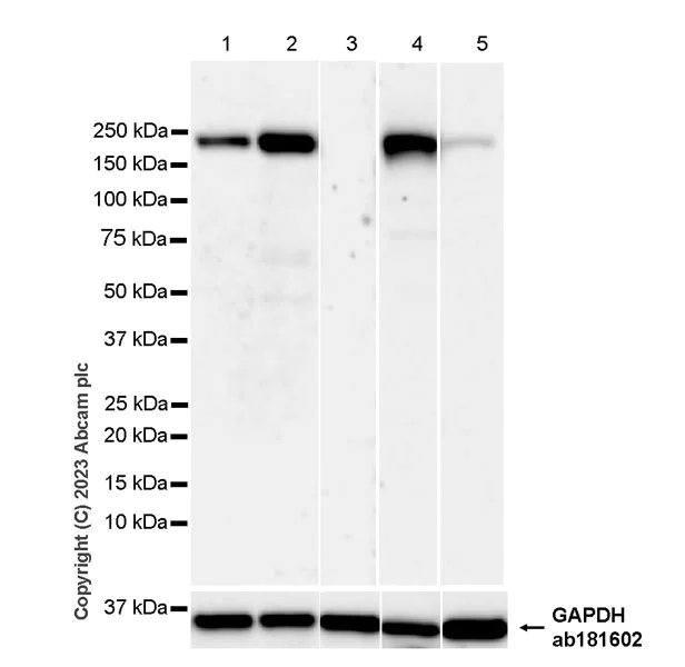
Western blot - Anti-Factor H antibody [EPR26491-30] (AB315879)
Blocking and diluting buffer and concentration : 5% NFDM/TBST.
Low expression : cerebellum.
Samples are non-boiled as boiling may cause protein aggregation.
Lanes 4-5 are applied with Goat Anti-Rabbit IgG, (H+L), Peroxidase conjugated (ab97051) at 1/100000 and lanes 1-3 are applied with Goat Anti-Rabbit IgG (HRP) with minimal cross-reactivity with human IgG at 1/2000.
In Western blot, Anti-GAPDH antibody [EPR16891] - Loading Control (ab181602) staining at 1/200000 dilution.
Exposure time : Lanes 1-3 : 203 seconds, lanes 4-5 : 37 seconds.
All lanes:
Western blot - Anti-Factor H antibody [EPR26491-30] (ab315879) at 1/1000 dilution
Lane 1:
Human kidney tissue lysate at 20 µg
Lane 2:
Human lung tissue lysate at 20 µg
Lane 3:
Human cerebellum tissue lysate at 20 µg
Lane 4:
Rat kidney tissue lysate at 20 µg
Lane 5:
Rat cerebellum tissue lysate at 20 µg
Secondary
Lanes 1 - 3:
Goat Anti-Rabbit IgG (HRP) with minimal cross-reactivity with human IgG at 1/2000 dilution
Lanes 4 - 5:
Western blot - Goat Anti-Rabbit IgG H&L (HRP) (<a href='/products/secondary-antibodies/goat-rabbit-igg-h-l-hrp-ab97051'>ab97051</a>) at 1/100000 dilution
Observed band size: 160 kDa,36 kDa
false
- WB
Supplier Data
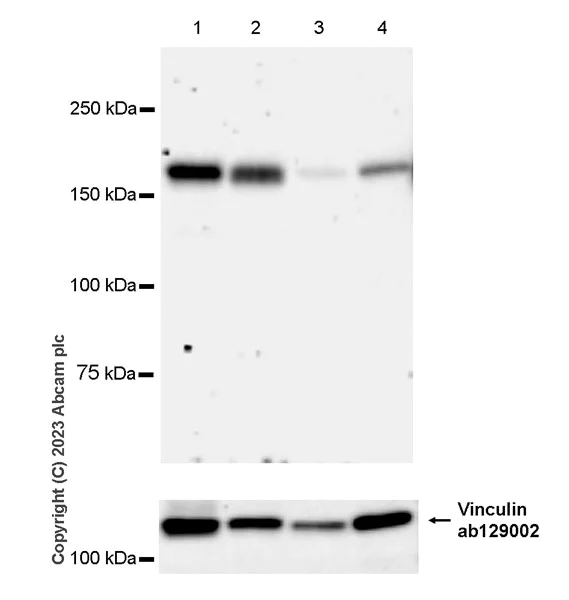
Western blot - Anti-Factor H antibody [EPR26491-30] (AB315879)
Blocking and diluting buffer and concentration : 5% NFDM/TBST.
Low expression : brain, spleen (PMID : 2533512).
Samples are non-boiled as boiling may cause protein aggregation.
In Western blot, Anti-Vinculin antibody [EPR8185] (ab129002) staining at 1/10000 dilution.
All lanes:
Western blot - Anti-Factor H antibody [EPR26491-30] (ab315879) at 1/1000 dilution
Lane 1:
Mouse kidney tissue lysate at 20 µg
Lane 2:
Mouse liver tissue lysate at 20 µg
Lane 3:
Mouse brain tissue lysate at 20 µg
Lane 4:
Mouse spleen tissue lysate at 20 µg
Secondary
All lanes:
Western blot - Goat Anti-Rabbit IgG H&L (HRP) (<a href='/products/secondary-antibodies/goat-rabbit-igg-h-l-hrp-ab97051'>ab97051</a>) at 1/100000 dilution
Observed band size: 160 kDa,124 kDa
false
Exposure time: 180s
- WB
Supplier Data
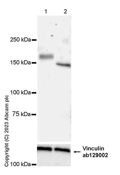
Western blot - Anti-Factor H antibody [EPR26491-30] (AB315879)
Factor H is a glycoprotein of approximately 160 kDa and detected as a 140-kDa band after treated with Peptide : N-glycosidase F (PNGase F).
Samples are non-boiled as boiling may cause protein aggregation.
In Western blot, Anti-Vinculin antibody [EPR8185] (ab129002) staining at 1/10000 dilution.
This blot was developed using a high sensitivity ECL substrate. The high-sensitivity ECL substrate used allows for the detection of proteins in the mid-femtogram range.
All lanes:
Western blot - Anti-Factor H antibody [EPR26491-30] (ab315879) at 1/1000 dilution
Lane 1:
Untreated Mouse liver tissue lysate at 20 µg
Lane 2:
Mouse liver tissue lysate treated with Peptide: N-glycosidase F (PNGase F) at 20 µg
Secondary
All lanes:
Western blot - Goat Anti-Rabbit IgG H&L (HRP) (<a href='/products/secondary-antibodies/goat-rabbit-igg-h-l-hrp-ab97051'>ab97051</a>) at 1/100000 dilution
Observed band size: 160 kDa,140 kDa,124 kDa
true
Exposure time: 180s
不同偶联物与剂型 (1)
-
Anti-Factor H antibody [EPR26491-30] - BSA and Azide free
搭配使用产品

Primary Antibodies
AB200999
20ul selling size|BOND RX™ Validated|RabMAb|Recombinant|KO Validated

- 4
- 9
Primary Antibodies
AB178846
Anti-Iba1 antibody [EPR16588] - Microglia marker
20ul selling size|BOND RX™ Validated|RabMAb|Recombinant|Lab Essentials

- 5
- 50


反应性数据
产品详情
Patented technology
Our RabMAb® technology is a patented hybridoma-based technology for making rabbit monoclonal antibodies. For details on our patents, please refer to RabMAb® patents.
What are the advantages of a recombinant monoclonal antibody?
This product is a recombinant monoclonal antibody, which offers several advantages including:
- - High batch-to-batch consistency and reproducibility
- - Improved sensitivity and specificity
- - Long-term security of supply
- - Animal-free batch production
For more information, read more on recombinant antibodies.
性能和储存信息
形式
纯化工艺
存储溶液
运输条件
推荐的短期储存时间
推荐的短期储存条件
推荐的长期储存条件
分装信息
储存信息
补充信息
This supplementary information is collated from multiple sources and compiled automatically.
Biological function summary
Factor H limits the activity of the complement system to prevent damage to host tissues. The protein exists in the plasma in a soluble form. It functions by recognizing host cell surfaces via specific markers avoiding inappropriate activation. Factor H belongs to a group of proteins which include other regulators of complement activation. These proteins maintain the balance between effective immune defense and protection of host tissue from excessive immune responses.
Pathways
Factor H is a part of the alternative complement pathway. This pathway is important for innate immune response involving proteins like factor P (properdin) which stabilizes C3 convertase. Factor H modulates these interactions to prevent unwarranted complement activity on host cells. Another related pathway is the classic complement pathway although factor H's involvement here is less direct since it primarily regulates the alternative pathway.
产品实验方案
- Visit the General protocols
- Visit the Troubleshooting
靶点信息
Abcam Product Promise
Please note: All products are 'FOR RESEARCH USE ONLY. NOT FOR USE IN DIAGNOSTIC OR THERAPEUTIC PROCEDURES'.
For licensing inquiries, please contact partnerships@abcam.com