Anti-Drosha抗体
Anti-Drosha antibody
4
(9 Reviews)
|
(100 Publications)
Rabbit Polyclonal Drosha antibody. Suitable for ICC, WB and reacts with Human, Mouse samples. Cited in 100 publications.
查看别名
RN3, RNASE3L, RNASEN, DROSHA, Ribonuclease 3, Protein Drosha, Ribonuclease III, p241, RNase III

- ICC
Unknown
Immunocytochemistry - Anti-Drosha antibody (AB12286)
ICC/IF image of ab12286 stained Hela cells. The cells were 4% PFA fixed (10 min) and then incubated in 1%BSA / 10% normal goat serum / 0.3M glycine in 0.1% PBS-Tween for 1h to permeabilise the cells and block non-specific protein-protein interactions. The cells were then incubated with the antibody (ab12886, 5μg/ml) overnight at +4°C. The secondary antibody (green) was DyLight® 488 goat anti-rabbit IgG - H&L, pre-adsorbed (ab96899) used at a 1/250 dilution for 1h. Alexa Fluor® 594 WGA was used to label plasma membranes (red) at a 1/200 dilution for 1h. DAPI was used to stain the cell nuclei (blue) at a concentration of 1.43μM.

- ICC
AbReview4126****
Immunocytochemistry - Anti-Drosha antibody (AB12286)
ab12286 at a 1/25 dilution staining human HeLa cells by immunocytochemistry. The antibody was incubated with the cells for 1 hour and then detected using a Cy3 conjugated donkey anti-rabbit polyclonal antibody.
This image is courtesy of an Abreview submitted on 7 February 2006.

- ICC
Collaborator
Immunocytochemistry - Anti-Drosha antibody (AB12286)
Human female amniocytes immunostained with ab12286 Drosha (FITC) (1/25 dilution). The DNA is labelled red with propidium iodide. This image was submitted as part of a review by Ahmad Khalil.
This image is courtesy of Ahmad M. Khalil and Daniel J. Driscoll, University of Florida College of Medicine Genetics Institute.

- ICC
Unknown
Immunocytochemistry - Anti-Drosha antibody (AB12286)
ICC/IF image of ab12286 stained HepG2 cells. The cells were 4% PFA fixed (10 min) and then incubated in 1%BSA / 10% normal goat serum / 0.3M glycine in 0.1% PBS-Tween for 1h to permeabilise the cells and block non-specific protein-protein interactions. The cells were then incubated with the antibody (ab12286, 1μg/ml) overnight at +4°C. The secondary antibody (green) was Alexa Fluor® 488 goat anti-rabbit IgG (H+L) used at a 1/1000 dilution for 1h. Alexa Fluor® 594 WGA was used to label plasma membranes (red) at a 1/200 dilution for 1h. DAPI was used to stain the cell nuclei (blue) at a concentration of 1.43μM.

- WB
Project
Western blot - Anti-Drosha antibody (AB12286)
Abcam recommends using milk as the blocking agent. Abcam welcomes customer feedback and would appreciate any comments regarding this product and the data presented above.
All lanes:
Western blot - Anti-Drosha antibody (ab12286) at 1 µg/mL
Lane 1:
HeLa (Human epithelial carcinoma cell line) Nuclear Lysate at 20 µg
Lane 2:
NIH 3T3 (Mouse embryonic fibroblast cell line) Nuclear Lysate at 20 µg
Secondary
All lanes:
Western blot - Goat Anti-Rabbit IgG H&L (HRP) preadsorbed (<a href='/products/secondary-antibodies/goat-rabbit-igg-h-l-hrp-preadsorbed-ab97080'>ab97080</a>) at 1/5000 dilution
Predicted band size: 159 kDa
Observed band size: 115 kDa,170 kDa
true
Exposure time: 12min

- WB
Project
Western blot - Anti-Drosha antibody (AB12286)
All lanes:
Western blot - Anti-Drosha antibody (ab12286) at 1 µg/mL
Lane 1:
HeLa (Human epithelial carcinoma cell line) Whole Cell Lysate at 20 µg
Lane 2:
HeLa (Human epithelial carcinoma cell line) Nuclear Lysate at 20 µg
Secondary
All lanes:
Goat polyclonal to Rabbit IgG (Alexa Fluor® 680) at 1/15000 dilution
Predicted band size: 159 kDa
Observed band size: 170 kDa
false

- WB
CiteAb
Western blot - Anti-Drosha antibody (AB12286)
Western Blotting using Anti-Drosha antibody, ab12286. Publication image from Zhang, Q. et al., 2015, Nat Commun, 25751603. Legend direct from paper.
ADAR1 mediates pri-miRNA processing(a) CLIP reads mapped to the mature, precursor and primary transcripts of miR-21-5p. Light and dark gray bars represent the relative locations of annotated mature and pre-miR-21. Stem-loop structure is shown for illustration purpose only that does not reflect the true structure of pre-miR-21. (b) Numbers of mature, pre-miR and pri-miR bound by ADAR1 and numbers of miRNAs with two or three forms bound by ADAR1 (shown as overlaps). Overlap p values between pri-miR and pre-miR : 0.024, between pri-miR and mature : 0.79 and between pre-miR and mature : 0.18, calculated using hypergeometric test and assuming a total of 410 miRNAs being expressed in U87MG cells (based on small RNA sequencing data). (c) Pri- (left panel) and mature miRNA expression (right panel) of endogenous miR-21-5p, miR-34a-5p, and miR-100-5p in U87MG cells as measured by RT-qPCR. Cells were transfected with the pEGFP-C1 vector (GFP), pEGFP-C1-ADAR1 vector (ADAR1), scrambled siRNA (siCtrl) or siRNA for ADAR1 (siADAR1) respectively. Results (mean and SD) from ≥4 biological replicates are shown. #p < 0.01, *p < 0.05 (Wilcoxon Rank-Sum test). (d) Expression change of miRNAs in U87MG cells with ADAR1 perturbation (KD or OE) relative to controls. Only miRNAs whose primary transcripts were associated with ADAR1 CLIP reads are included. Filled circles represent miRNAs with significantly altered expression in KD or OE. If the significance was observed in both experiments or if no significance was found in either experiment, the average expression change is shown. If only one experiment led to a significant change, the value of expression change in this experiment is shown. Two miRNAs were excluded because they demonstrated significant changes in both experiments, but in conflicting directions. (e) Cumulative distribution functions of log2-fold changes (LFCs) of miRNAs bound by ADAR1 in the primary form in U87MG cells upon OE of wildtype ADAR1, E912A or EAA mutants, compared to controls. The LFC values represent log2(OE/ctrls) which were further normalized by spike-in controls to account for technical variations across experiments. (f) ADAR1 associates with both DROSHA and DGCR8. Co-immunoprecipitation (Co-IP) experiments were performed using DROSHA antibody (ab12286), DGCR8 antibody (ab90579), ADAR1 antibody (D-8), or corresponding rabbit (r) or mouse (m) isotype IgG in HeLa cells. HeLa cells were used since DROSHA and DGCR8 expression is relatively higher in HeLa than in U87MG cells. IP samples were immunoblotted (IB) using ADAR1 antibody (15.8.6), DROSHA antibody (ab12286) and DGCR8 antibody (ab90579) to detect the corresponding antigens. ADAR1 interacts with both DROSHA and DGCR8, reciprocally but not the corresponding IgG isotype control. RNase A treatment does not significantly impair the interactions.
false
- WB
CiteAb
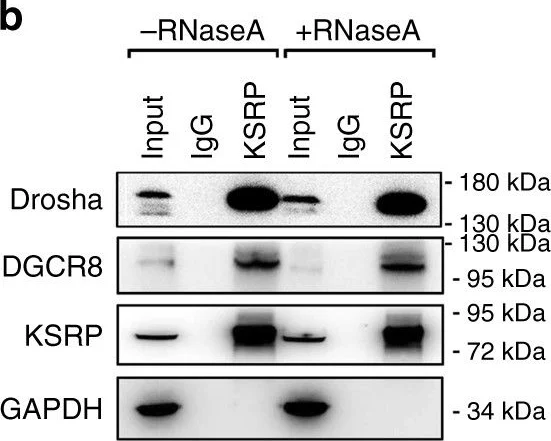
Western blot - Anti-Drosha antibody (AB12286)
Western Blotting using Anti-Drosha antibody, ab12286. Publication image from Wang, F. et al., 2017, Nat Commun, 29127290. Legend direct from paper.
KSRP promotes the processing of pri-129-1 in vitro. a Schematic representation of the involvement of KSRP in the pri-129-1 processing model. b As shown in the co-IP analysis, KSRP interacts with the Drosha-DGCR8 complex in THP-1 cells in an RNA-independent manner. This experiment was performed by incubating the KSRP immunoprecipitates with or without RNAse A (0.1 mg/mL) at 37 °C for 20 min. c Agarose gel electrophoresis showing the results of the RIP-PCR of pri-129-1, pri-23b and pri-let7a was performed by immunoprecipitating the complexes in KSRP knockdown or control 293T cells with Drosha and DGCR8 antibodies. d Representative images for in situ proximity ligation assay (PLA) of KSRP with Drosha or DGCR8 in 293T cells (Scale bar, 10 µm; Original magnification, 630x). e Quantification of PLA results (n ≥ 3; cell numbers > 100 per experiment) were shown. f In vitro pri-129-1 processing reactions used isotope-labeled wild-type pri-129-1 (129_WT) or pri-129-1 with KSRP-binding site mutants (all four sites mutant, 129_Mut) as the substrate and were then pre-incubated with HeLa NEs and various amounts of purified KSRP protein, as described in the Methods. g Rescue of the KSRP levels in NEs after KSRP depletion with an anti-KSRP antibody further promotes pri-129-1 processing in the in vitro processing assay, as indicated in f. h Quantitation of the in vitro processing assay shown in g. Similar significant differences (p-value < 0.05, two-tailed Student’s t-test) were obtained between the KSRP depletion group and the KSRP rescue #1 or #2 groups. Three technical replicates from a single experiment. Data are shown as means±s.d. ***P < 0.001, Student’s t-test
false
替代产品


Primary Antibodies
AB227460
Anti-Drosha antibody [EPR12794] - BSA and Azide free
RabMAb|Recombinant|KO Validated

- 0
- 0

搭配使用产品
Primary Antibodies
AB191875
Anti-DGCR8 antibody [EPR18757]
20ul selling size|KO Validated|RabMAb|Recombinant

- 5
- 2
Primary Antibodies
AB259327
Anti-Dicer antibody [EPR24104-105]
BOND RX™ Validated|20ul selling size|RabMAb|Recombinant|KO Validated

- 0
- 0
Primary Antibodies
AB183734
RabMAb|Recombinant|Lab Essentials|20ul selling size

- 5
- 24

反应性数据
产品详情
性能和储存信息
形式
纯化工艺
存储溶液
运输条件
推荐的短期储存时间
推荐的短期储存条件
推荐的长期储存条件
分装信息
储存信息
产品实验方案
- Visit the General protocols
- Visit the Troubleshooting
靶点信息
文献 (100)
Recent publications for all applications. Explore the full list and refine your search
Biochimica et biophysica acta. Gene regulatory mechanisms 1867:195065 PubMed39455000
2024
Applications
Unspecified application
Species
Unspecified reactive species
Science advances 9:eadf0597 PubMed36763664
2023
Applications
Unspecified application
Species
Unspecified reactive species
Viruses 15: PubMed36851522
2023
Applications
Unspecified application
Species
Unspecified reactive species
Aging 14:6520-6536 PubMed35951366
2022
Applications
Unspecified application
Species
Unspecified reactive species
FASEB journal : official publication of the Federation of American Societies for Experimental Biology 36:e22412 PubMed35713587
2022
Applications
Unspecified application
Species
Unspecified reactive species
Journal of translational medicine 20:207 PubMed35545774
2022
Applications
Unspecified application
Species
Unspecified reactive species
Cancer science 113:1601-1612 PubMed35253323
2022
Applications
Unspecified application
Species
Unspecified reactive species
MicroRNA (Shariqah, United Arab Emirates) 11:36-44 PubMed35184719
2022
Applications
Unspecified application
Species
Unspecified reactive species
Cell death and differentiation 28:2708-2727 PubMed33875796
2021
Applications
Unspecified application
Species
Unspecified reactive species
Nucleic acids research 49:2522-2536 PubMed33561291
2021
Applications
Unspecified application
Species
Unspecified reactive species
Abcam Product Promise
Please note: All products are 'FOR RESEARCH USE ONLY. NOT FOR USE IN DIAGNOSTIC OR THERAPEUTIC PROCEDURES'.
For licensing inquiries, please contact partnerships@abcam.com