Anti-c-Fos 抗体 [N486/32]
Anti-c-Fos antibody [N486/32]
- BOND RX™ Validated
- Recombinant
- Lab Essentials
- KO Validated
- 了解详情
Be the first to review this product! Submit a review
|
(5 Publications)
Mouse Recombinant Monoclonal c-Fos antibody. Suitable for WB, IHC-P, ICC/IF and reacts with Human, Mouse, Rat samples. Cited in 5 publications.
查看别名
G0S7, FOS, Protein c-Fos, Cellular oncogene fos, G0/G1 switch regulatory protein 7, Proto-oncogene c-Fos, Transcription factor AP-1 subunit c-Fos

- IHC-P
Supplier Data
Immunohistochemistry (Formalin/PFA-fixed paraffin-embedded sections) - Anti-c-Fos antibody [N486/32] (AB302667)
Immunohistochemical analysis of paraffin-embedded Human cervical cancer tissue labeling c-Fos with ab302667 at 1/500 (1.676 ug/ml) followed by a ready to use LeicaDS9800 (Bond™ Polymer Refine Detection) was used. Nuclear staining on human cervical cancer.The section was incubated with ab302667 for 30 mins at room temperature, followed by anti-mouse IgG2a antibody (610-4141) for 8 mins during the LeicaDS9800 kit staining procedure. The immunostaining was performed on a Leica Biosystems BOND® RX instrument Counterstained with Hematoxylin. Secondary antibody only control : Secondary antibody is a ready to use LeicaDS9800 (Bond™ Polymer Refine Detection) was used. Heat mediated antigen retrieval with Citrate buffer (pH 6.0, epitope retrieval solution 1) for 20 mins

- IHC-P
Supplier Data
Immunohistochemistry (Formalin/PFA-fixed paraffin-embedded sections) - Anti-c-Fos antibody [N486/32] (AB302667)
Immunohistochemical analysis of paraffin-embedded Human colon tissue labeling c-Fos with ab302667 at 1/500 (1.676 ug/ml) followed by a ready to use LeicaDS9800 (Bond Polymer Refine Detection) was used. Nuclear staining on human colon.The section was incubated with ab302667 for 30 mins at room temperature, followed by anti-mouse IgG2a antibody (610-4141) for 8 mins during the LeicaDS9800 kit staining procedure.The immunostaining was performed on a Leica Biosystems BOND® RX instrument Counterstained with Hematoxylin. Secondary antibody only control : Secondary antibody is a ready to use LeicaDS9800 (Bond Polymer Refine Detection) was used. Heat mediated antigen retrieval with Citrate buffer (pH 6.0, epitope retrieval solution 1) for 20 mins

- ICC/IF
Supplier Data
Immunocytochemistry/ Immunofluorescence - Anti-c-Fos antibody [N486/32] (AB302667)
Immunofluorescent analysis of 4% Paraformaldehyde-fixed, 0.1% TritonX-100 permeabilized HeLa (human cervix adenocarcinoma epithelial cell) cells labelling c-Fos with ab302667 at 1/250 (3.4 ug/ml) dilution, followed by ab150117 Goat Anti-Mouse IgG H&L (Alexa Fluor® 488) preadsorbed antibody at 1/1000 2 ug/ml dilution (Green). Confocal image showing increased nuclear staining in HeLa cells after starvation 16 hours then treated with TPA (200 nM) for 4 h is observed. ab179513 Anti-beta Tubulin rabbit monoclonal antibody was used to counterstain tubulin at 1/200 10ug/ml dilution (Red). The Nuclear counterstain was DAPI (Blue). Secondary antibody only control : Secondary antibody is ab150117 Goat Anti-Mouse IgG H&L (Alexa Fluor® 488) preadsorbed at 1/1000 2 ug/ml dilution.

- ICC/IF
Supplier Data
Immunocytochemistry/ Immunofluorescence - Anti-c-Fos antibody [N486/32] (AB302667)
Immunofluorescent analysis of 4% Paraformaldehyde-fixed, 0.1% TritonX-100 permeabilized NIH/3T3 (mouse embryonic fibroblast) cells labelling c-Fos with ab302667 at 1/250 (3.4 ug/ml) dilution, followed by ab150117 Goat Anti-Mouse IgG H&L (Alexa Fluor® 488) preadsorbed antibody at 1/1000 2 ug/ml dilution (Green). Confocal image showing increased nuclear staining in NIH/3T3 cells after starvation 16 hours then treated with TPA (200 nM) and MG-132(10 µM) for 4 h is observed. ab179513 Anti-beta Tubulin rabbit monoclonal antibody was used to counterstain tubulin at 1/200 10ug/ml dilution (Red). The Nuclear counterstain was DAPI (Blue). Secondary antibody only control : Secondary antibody is ab150117 Goat Anti-Mouse IgG H&L (Alexa Fluor® 488) preadsorbed at 1/1000 2 ug/ml dilution.

- IHC-P
Supplier Data
Immunohistochemistry (Formalin/PFA-fixed paraffin-embedded sections) - Anti-c-Fos antibody [N486/32] (AB302667)
Immunohistochemical analysis of paraffin-embedded Mouse cerebrum tissue labeling c-Fos with ab302667 at 1/2000 (0.419 ug/ml) followed by a ready to use LeicaDS9800 (Bond Polymer Refine Detection) was used. Nuclear staining on mouse cerebrum.The section was incubated with ab302667 for 30 mins at room temperature, followed by anti-mouse IgG2a antibody (610-4141) for 8 mins during the LeicaDS9800 kit staining procedure.The immunostaining was performed on a Leica Biosystems BOND® RX instrument Counterstained with Hematoxylin. Secondary antibody only control : Secondary antibody is a ready to use LeicaDS9800 (Bond Polymer Refine Detection) was used. Heat mediated antigen retrieval with Citrate buffer (pH 6.0, epitope retrieval solution 1) for 20 mins

- IHC-P
Supplier Data
Immunohistochemistry (Formalin/PFA-fixed paraffin-embedded sections) - Anti-c-Fos antibody [N486/32] (AB302667)
Immunohistochemical analysis of paraffin-embedded Rat cerebrum tissue labeling c-Fos with ab302667 at 1/1000 (0.838 ug/ml) followed by a ready to use LeicaDS9800 (Bond Polymer Refine Detection) was used. Nuclear staining on rat cerebrum.The section was incubated with ab302667 for 30 mins at room temperature, followed by anti-mouse IgG2a antibody (610-4141) for 8 mins during the LeicaDS9800 kit staining procedure.The immunostaining was performed on a Leica Biosystems BOND® RX instrument Counterstained with Hematoxylin. Secondary antibody only control : Secondary antibody is a ready to use LeicaDS9800 (Bond Polymer Refine Detection) was used. Heat mediated antigen retrieval with Citrate buffer (pH 6.0, epitope retrieval solution 1) for 20 mins

- WB
Supplier Data
Western blot - Anti-c-Fos antibody [N486/32] (AB302667)
Data of BM-Recombinant Anti-c-Fos antibody [EPR20769] (ab214672) was in the internal.Blocking and diluting buffer and concentration : 5% NFDM/TBST The band(s) beneath the target band are likely to be degraded target fragments (PMID : 9737957 and PMID : 20498278). Exposure time : Lanes 1-2 : 26 seconds Lanes 3-4 : 81 seconds Lanes 5-6 : 136 seconds
All lanes:
Western blot - Anti-c-Fos antibody [N486/32] (ab302667) at 1/1000 dilution
Lane 1:
HeLa (human cervix adenocarcinoma epithelial cell) starved overnight, whole cell lysate 20 µg
Lane 2:
HeLa starved overnight, then treated with 200nM PMA (<a href='/products/biochemicals/phorbol-12-myristate-13-acetate-pma-pkc-activator-ab120297'>ab120297</a>) for 4 hours, whole cell lysate 20 µg
Lane 3:
NIH/3T3 (mouse embryonic fibroblast) starved overnight, whole cell lysate 20 µg
Lane 4:
NIH/3T3 starved overnight, then treated with 200nM PMA (<a href='/products/biochemicals/phorbol-12-myristate-13-acetate-pma-pkc-activator-ab120297'>ab120297</a>) and 10uM MG-132 (<a href='/products/biochemicals/mg-132-proteasome-inhibitor-ab141003'>ab141003</a>) for 4 hours, whole cell lysate for 4 hours, whole cell lysate 20 µg
Lane 5:
PC-12 (rat adrenal gland pheochromocytoma) starved overnight, whole cell lysate 20 µg
Lane 6:
PC-12 starved overnight, then treated with /ml NGF (<a href='/products/proteins-peptides/recombinant-human-ngf-protein-active-ab9796'>ab9796</a>) and 10uM MG-132 (<a href='/products/biochemicals/mg-132-proteasome-inhibitor-ab141003'>ab141003</a>) for 2 hours, whole cell lysate 20 µg
Secondary
All lanes:
Peroxidase-Conjugated Goat anti-Mouse IgG (H+L) at 1/10000 dilution
Observed band size: 37 kDa,55 kDa
false
Exposure time: 26s
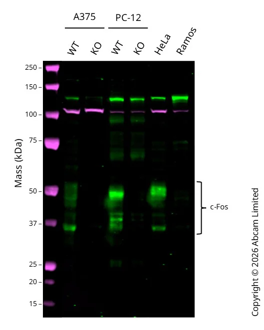
- WB
Lab
Western blot - Anti-c-Fos antibody [N486/32] (AB302667)
Western blot : Anti-c-Fos antibody [N486/32] ab302667 staining at 1/1000 dilution, shown in green; Rabbit anti-ACTN4 ab108198 loading control staining at 1/20000 dilution, shown in magenta. A band was observed at 35-50 kDa in Wild-type A375 cell lysates with no signal observed at this size in FOS knockout A375 cell line. To generate this image, samples were run on an SDS-PAGE gel then transferred onto a nitrocellulose membrane. Membranes were blocked in 3 % Milk in TBS-0.1 % Tween® 20 (TBS-T) before incubation with primary antibodies overnight at 4 °C. Blots were washed four times in TBS-T, incubated with secondary antibodies for 1 h at room temperature, washed again four times then imaged. Secondary antibodies used were Goat anti-Mouse 800CW and Goat anti-Rabbit 680RD at 1/20,000 dilution.
All lanes:
Western blot - Anti-c-Fos antibody [N486/32] (ab302667) at 1/1000 dilution
Lane 1:
Wild-type A375 cell lysates at 20 µg
Lane 2:
FOS knockout A375 cell lysates at 20 µg
Lane 3:
Wild-type PC-12 ab279978 cell lysates at 20 µg
Lane 4:
Western blot - Rat FOS (c-Fos) knockout PC-12 cell line (ab281615) at 20 µg
Lane 5:
HeLa cell lysates at 20 µg
Lane 6:
Ramos cell lysates at 20 µg
Secondary
All lanes:
Goat anti-Mouse 800CW & Goat anti-Rabbit 680RD at 1/20000 dilution
Predicted band size: 41 kDa
Observed band size: 35-50 kDa
false
不同偶联物与剂型 (1)
-
Anti-c-Fos antibody [N486/32] - BSA and Azide free
搭配使用产品

Primary Antibodies
AB183734
RabMAb|Recombinant|Lab Essentials|20ul selling size

- 5
- 24
Primary Antibodies
AB177487
Anti-NeuN antibody [EPR12763] - Neuronal Marker
BOND RX™ Validated|RabMAb|Recombinant|Lab Essentials|20ul selling size|Advanced Validation

- 5
- 75
Primary Antibodies
AB178846
Anti-Iba1 antibody [EPR16588] - Microglia marker
20ul selling size|BOND RX™ Validated|RabMAb|Recombinant|Lab Essentials

- 5
- 50
Primary Antibodies
AB278054
Anti-GFAP antibody [RM1003] - Astrocyte Marker
BOND RX™ Validated|20ul selling size|RabMAb|Recombinant

- 0
- 0
反应性数据
产品详情
What are the advantages of a recombinant monoclonal antibody?
This product is a recombinant monoclonal antibody, which offers several advantages including:
- - High batch-to-batch consistency and reproducibility
- - Improved sensitivity and specificity
- - Long-term security of supply
- - Animal-free batch production
For more information, read more on recombinant antibodies.
Want a custom formulation?
This antibody clone is manufactured by Abcam. If you require a custom buffer formulation or conjugation for your experiments, please contact orders@abcam.com
性能和储存信息
形式
纯化工艺
存储溶液
运输条件
推荐的短期储存时间
推荐的短期储存条件
推荐的长期储存条件
分装信息
储存信息
补充信息
This supplementary information is collated from multiple sources and compiled automatically.
Biological function summary
C-Fos acts as part of the Activator Protein-1 (AP-1) complex a group of DNA-binding proteins that regulate gene transcription. This complex forms when c-Fos and Jun proteins dimerize. The AP-1 complex controls several cellular processes such as proliferation differentiation and apoptosis. Due to its participation in these vital processes c-Fos influences cellular responses to external stimuli and can affect how cells behave under stress.
Pathways
C-Fos significantly contributes to the MAPK/ERK pathway which plays a role in cell growth and differentiation. It partners with Jun proteins to form functional transcription factors within the MAPK signaling cascade. Additionally c-Fos is involved in the JNK signaling pathway linking it to stress responses and apoptosis. These pathways highlight the partnerships between c-Fos and other proteins like ERK and JNK indicating their intertwined roles in cellular signaling networks.
产品实验方案
- Visit the General protocols
- Visit the Troubleshooting
靶点信息
文献 (5)
Recent publications for all applications. Explore the full list and refine your search
Nature communications 16:8870 PubMed41053178
2025
Applications
Unspecified application
Species
Unspecified reactive species
Nature communications 16:4437 PubMed40360512
2025
Applications
Unspecified application
Species
Unspecified reactive species
Advanced science (Weinheim, Baden-Wurttemberg, Germany) 12:e2415601 PubMed40245175
2025
Applications
Unspecified application
Species
Unspecified reactive species
Neuropsychopharmacology : official publication of the American College of Neuropsychopharmacology 49:1468-1480 PubMed38734818
2024
Applications
Unspecified application
Species
Unspecified reactive species
Stem cells translational medicine 12:307-321 PubMed37010483
2023
Applications
Unspecified application
Species
Unspecified reactive species
Abcam Product Promise
Please note: All products are 'FOR RESEARCH USE ONLY. NOT FOR USE IN DIAGNOSTIC OR THERAPEUTIC PROCEDURES'.
For licensing inquiries, please contact partnerships@abcam.com