重组Anti-ARMET/ARP抗体[EPR29115-74]
Anti-ARMET/ARP antibody [EPR29115-74]
- BOND RX™ Validated
- 20ul selling size
- Recombinant
- Advanced Validation
- RabMAb
- 了解详情
Be the first to review this product! Submit a review
|
(1 Publication)
Rabbit Recombinant Monoclonal ARMET/ARP antibody. Suitable for WB, Flow Cyt (Intra), IP, IHC-P, ICC/IF, mIHC and reacts with Recombinant fragment - Mouse, Human, Mouse, Rat samples. Cited in 1 publication.
查看别名
Armet, Manf, Mesencephalic astrocyte-derived neurotrophic factor, Arginine-rich protein, Protein ARMET

- IHC-P
Supplier Data
Immunohistochemistry (Formalin/PFA-fixed paraffin-embedded sections) - Anti-ARMET/ARP antibody [EPR29115-74] (AB316935)
Immunohistochemical analysis of paraffin-embedded Human skeletal muscle tissue labeling ARMET/ARP with ab316935 at 1/2000 (0.253 ug/ml) dilution, followed by a ready to use LeicaDS9800 (Bond™ Polymer Refine Detection).
Low expression : no staining on human skeletal muscle. The section was incubated with ab316935 for 30 mins at room temperature.
The immunostaining was performed on a Leica Biosystems BOND® RX instrument.
Counterstained with Hematoxylin.
Secondary antibody only control : Secondary antibody is a ready to use LeicaDS9800 (Bond™ Polymer Refine Detection).
Heat mediated antigen retrieval was performed with Tris-EDTA buffer (pH 9.0, Epitope Retrieval Solution2) for 20 mins.

- mIHC
Supplier Data
Multiplex immunohistochemistry - Anti-ARMET/ARP antibody [EPR29115-74] (AB316935)
Multiplex immunohistochemical analysis of formalin/PFA-fixed paraffin-embedded human cerebrum tissue section labelling Noradrenaline transporter with ab254361 at a 1/100 dilution (B), GPCR GPR17 with ab314307 at a 1/500 dilution (C), and ARMET/ARP with ab316935 at a 1/1000 dilution (D). Nuclear DNA was labeled with DAPI (shown in blue).
Panel A : merged staining of anti-Noradrenaline transporter (magenta; Opal™ 690), anti-GPCR GPR17 (green; Opal™ 520) and anti-ARMET/ARP (gray; Opal™ 570) on human cerebrum.
Panel B : anti-Noradrenaline transporter staining nerves in human cerebrum.
Panel C : anti-GPCR GPR17 staining oligodendrocytes in human cerebrum.
Panel D : anti-ARMET/ARP staining neurons in human cerebrum.
Heat mediated antigen retrieval with Tris-EDTA buffer (pH 9.0, epitope retrieval solution2) for 20 mins.
The section was incubated in three rounds of staining : in the order of ab254361, ab314307 and ab316935 for 30 mins at room temperature. Each round was followed by a separate fluorescent tyramide signal amplification system.
The immunostaining was performed on a Leica Biosystems BOND® RX instrument with an Opal™ 4-color kit. Image acquisition was performed with Leica SP8 confocal microscope.

- IHC-P
Supplier Data
Immunohistochemistry (Formalin/PFA-fixed paraffin-embedded sections) - Anti-ARMET/ARP antibody [EPR29115-74] (AB316935)
Immunohistochemical analysis of paraffin-embedded Human cerebrum tissue labeling ARMET/ARP with ab316935 at 1/2000 (0.253 ug/ml) dilution, followed by a ready to use LeicaDS9800 (Bond™ Polymer Refine Detection).
Positive staining on neurons of human cerebrum. The section was incubated with ab316935 for 30 mins at room temperature.
The immunostaining was performed on a Leica Biosystems BOND® RX instrument.
Counterstained with Hematoxylin.
Secondary antibody only control : Secondary antibody is a ready to use LeicaDS9800 (Bond™ Polymer Refine Detection).
Heat mediated antigen retrieval was performed with Tris-EDTA buffer (pH 9.0, Epitope Retrieval Solution2) for 20 mins.

- mIHC
Supplier Data
Multiplex immunohistochemistry - Anti-ARMET/ARP antibody [EPR29115-74] (AB316935)
Multiplex immunohistochemical analysis of formalin/PFA-fixed paraffin-embedded mouse cerebellum tissue section labelling Noradrenaline transporter with ab254361 at a 1/100 dilution (B), GPCR GPR17 with ab314307 at a 1/2000 dilution (C), and ARMET/ARP with ab316935 at a 1/20000 dilution (D). Nuclear DNA was labeled with DAPI (shown in blue).
Panel A : merged staining of anti-Noradrenaline transporter (magenta; Opal™ 690), anti-GPCR GPR17 (green; Opal™ 520) and anti-ARMET/ARP (gray; Opal™ 570) on mouse cerebellum.
Panel B : anti-Noradrenaline transporter staining nerves in mouse cerebellum.
Panel C : anti-GPCR GPR17 staining oligodendrocytes in mouse cerebellum.
Panel D : anti-ARMET/ARP staining neurons in mouse cerebellum.
Heat mediated antigen retrieval with Tris-EDTA buffer (pH 9.0, epitope retrieval solution2) for 20 mins.
The section was incubated in three rounds of staining : in the order of ab254361, ab314307 and ab316935 for 30 mins at room temperature. Each round was followed by a separate fluorescent tyramide signal amplification system.
The immunostaining was performed on a Leica Biosystems BOND® RX instrument with an Opal™ 4-color kit. Image acquisition was performed with Leica SP8 confocal microscope.

- mIHC
Supplier Data
Multiplex immunohistochemistry - Anti-ARMET/ARP antibody [EPR29115-74] (AB316935)
Multiplex immunohistochemistry analysis of formalin/PFA-fixed paraffin-embedded rat spinal cord tissue section labelling GPCR GPR17 with ab314307 at 1/2000 dilution (B), TAC1 with ab307138 at 1/500 dilution (C), and ARMET/ARP with ab316935 at 1/20000 dilution (D). Nuclear DNA was labeled with DAPI (shown in blue). Heat mediated antigen retrieval with Tris-EDTA buffer (pH 9.0, epitope retrieval solution2) for 20 mins.
Panel A : merged staining of anti-GPCR GPR17 (magenta; Opal™ 690), anti-TAC1 (green; Opal™ 520) and anti-ARMET/ARP (yellow; Opal™ 570) on rat spinal cord.
Panel B : anti-GPCR GPR17 staining oligodendrocytes in rat spinal cord.
Panel C : anti-TAC1 staining posterior horns (sensory) in rat spinal cord.
Panel D : anti-ARMET/ARP staining neurons in rat spinal cord.
The section was incubated in three rounds of staining : in the order of ab314307, ab307138 and ab316935 for 30 mins at room temperature. Each round was followed by a separate fluorescent tyramide signal amplification system.
The immunostaining was performed on a Leica Biosystems BOND® RX instrument with an Opal™ 4-color kit. Image acquisition was performed with Leica SP8 confocal microscope.

- mIHC
Supplier Data
Multiplex immunohistochemistry - Anti-ARMET/ARP antibody [EPR29115-74] (AB316935)
Multiplex immunohistochemistry analysis of formalin/PFA-fixed paraffin-embedded mouse spinal cord tissue section labelling GPCR GPR17 with ab314307 at 1/2000 dilution (B), TAC1 with ab307138 at 1/500 dilution (C), and ARMET/ARP with ab316935 at 1/20000 (D). Nuclear DNA was labeled with DAPI (shown in blue). Heat mediated antigen retrieval with Tris-EDTA buffer (pH 9.0, epitope retrieval solution2) for 20 mins.
Panel A : merged staining of anti-GPCR GPR17 (magenta; Opal™ 690), anti-TAC1 (green; Opal™ 520) and anti-ARMET/ARP (yellow; Opal™ 570) on mouse spinal cord.
Panel B : anti-GPCR GPR17 staining oligodendrocytes in mouse spinal cord.
Panel C : anti-TAC1 staining posterior horns (sensory) in mouse spinal cord.
Panel D : anti-ARMET/ARP staining neurons in mouse spinal cord.
The section was incubated in three rounds of staining : in the order of ab314307, ab307138 and ab316935 for 30 mins at room temperature. Each round was followed by a separate fluorescent tyramide signal amplification system.
The immunostaining was performed on a Leica Biosystems BOND® RX instrument with an Opal™ 4-color kit. Image acquisition was performed with Leica SP8 confocal microscope.

- mIHC
Supplier Data
Multiplex immunohistochemistry - Anti-ARMET/ARP antibody [EPR29115-74] (AB316935)
Multiplex immunohistochemical analysis of formalin/PFA-fixed paraffin-embedded mouse cerebrum tissue section labelling Noradrenaline transporter with ab254361 at a 1/100 dilution (B), GPCR GPR17 with ab314307 at a 1/2000 dilution (C), and ARMET/ARP with ab316935 at a 1/20000 dilution (D). Nuclear DNA was labeled with DAPI (shown in blue).
Panel A : merged staining of anti-Noradrenaline transporter (magenta; Opal™ 690), anti-GPCR GPR17 (green; Opal™ 520) and anti-ARMET/ARP (gray; Opal™ 570) on mouse cerebrum.
Panel B : anti-Noradrenaline transporter staining nerves in mouse cerebrum.
Panel C : anti-GPCR GPR17 staining oligodendrocytes in mouse cerebrum.
Panel D : anti-ARMET/ARP staining neurons in mouse cerebrum.
Heat mediated antigen retrieval with Tris-EDTA buffer (pH 9.0, epitope retrieval solution2) for 20 mins.
The section was incubated in three rounds of staining : in the order of ab254361, ab314307 and ab316935 for 30 mins at room temperature. Each round was followed by a separate fluorescent tyramide signal amplification system.
The immunostaining was performed on a Leica Biosystems BOND® RX instrument with an Opal™ 4-color kit. Image acquisition was performed with Leica SP8 confocal microscope.

- mIHC
Supplier Data
Multiplex immunohistochemistry - Anti-ARMET/ARP antibody [EPR29115-74] (AB316935)
Multiplex immunohistochemical analysis of formalin/PFA-fixed paraffin-embedded rat cerebellum tissue section labelling Noradrenaline transporter with ab254361 at a 1/100 dilution (B), GPCR GPR17 with ab314307 at a 1/2000 dilution (C), and ARMET/ARP with ab316935 at a 1/20000 dilution (D). Nuclear DNA was labeled with DAPI (shown in blue).
Panel A : merged staining of anti-Noradrenaline transporter (magenta; Opal™ 690), anti-GPCR GPR17 (green; Opal™ 520) and anti-ARMET/ARP (gray; Opal™ 570) on rat cerebellum.
Panel B : anti-Noradrenaline transporter staining nerves in rat cerebellum.
Panel C : anti-GPCR GPR17 staining oligodendrocytes in rat cerebellum.
Panel D : anti-ARMET/ARP staining neurons in rat cerebellum.
Heat mediated antigen retrieval with Tris-EDTA buffer (pH 9.0, epitope retrieval solution2) for 20 mins.
The section was incubated in three rounds of staining : in the order of ab254361, ab314307 and ab316935 for 30 mins at room temperature. Each round was followed by a separate fluorescent tyramide signal amplification system.
The immunostaining was performed on a Leica Biosystems BOND® RX instrument with an Opal™ 4-color kit. Image acquisition was performed with Leica SP8 confocal microscope.

- mIHC
Supplier Data
Multiplex immunohistochemistry - Anti-ARMET/ARP antibody [EPR29115-74] (AB316935)
Multiplex immunohistochemical analysis of formalin/PFA-fixed paraffin-embedded rat cerebrum tissue section labelling Noradrenaline transporter with ab254361 at a 1/100 dilution (B), GPCR GPR17 with ab314307 at a 1/2000 dilution (C), and ARMET/ARP with ab316935 at a 1/20000 dilution (D). Nuclear DNA was labeled with DAPI (shown in blue).
Panel A : merged staining of anti-Noradrenaline transporter (magenta; Opal™ 690), anti-GPCR GPR17 (green; Opal™ 520) and anti-ARMET/ARP (gray; Opal™ 570) on rat cerebrum.
Panel B : anti-Noradrenaline transporter staining nerves in rat cerebrum.
Panel C : anti-GPCR GPR17 staining oligodendrocytes in rat cerebrum.
Panel D : anti-ARMET/ARP staining neurons in rat cerebrum.
Heat mediated antigen retrieval with Tris-EDTA buffer (pH 9.0, epitope retrieval solution2) for 20 mins.
The section was incubated in three rounds of staining : in the order of ab254361, ab314307 and ab316935 for 30 mins at room temperature. Each round was followed by a separate fluorescent tyramide signal amplification system.
The immunostaining was performed on a Leica Biosystems BOND® RX instrument with an Opal™ 4-color kit. Image acquisition was performed with Leica SP8 confocal microscope.

- IHC-P
Supplier Data
Immunohistochemistry (Formalin/PFA-fixed paraffin-embedded sections) - Anti-ARMET/ARP antibody [EPR29115-74] (AB316935)
Immunohistochemical analysis of paraffin-embedded Mouse skeletal muscle tissue labeling ARMET/ARP with ab316935 at 1/10000 (0.051 ug/ml) dilution, followed by a ready to use LeicaDS9800 (Bond™ Polymer Refine Detection).
Low expression : no staining on mouse skeletal muscle. The section was incubated with ab316935 for 30 mins at room temperature.
The immunostaining was performed on a Leica Biosystems BOND® RX instrument.
Counterstained with Hematoxylin.
Secondary antibody only control : Secondary antibody is a ready to use LeicaDS9800 (Bond™ Polymer Refine Detection).
Heat mediated antigen retrieval was performed with Tris-EDTA buffer (pH 9.0, Epitope Retrieval Solution2) for 20 mins.

- IHC-P
Supplier Data
Immunohistochemistry (Formalin/PFA-fixed paraffin-embedded sections) - Anti-ARMET/ARP antibody [EPR29115-74] (AB316935)
Immunohistochemical analysis of paraffin-embedded Mouse P5 cerebrum tissue labeling ARMET/ARP with ab316935 at 1/10000 (0.051 ug/ml) dilution, followed by a ready to use LeicaDS9800 (Bond™ Polymer Refine Detection).
Positive staining on neurons of mouse cerebrum (PMID : 18718866). The section was incubated with ab316935 for 30 mins at room temperature.
The immunostaining was performed on a Leica Biosystems BOND® RX instrument.
Counterstained with Hematoxylin.
Secondary antibody only control : Secondary antibody is a ready to use LeicaDS9800 (Bond™ Polymer Refine Detection).
Heat mediated antigen retrieval was performed with Tris-EDTA buffer (pH 9.0, Epitope Retrieval Solution2) for 20 mins.

- IHC-P
Supplier Data
Immunohistochemistry (Formalin/PFA-fixed paraffin-embedded sections) - Anti-ARMET/ARP antibody [EPR29115-74] (AB316935)
Immunohistochemical analysis of paraffin-embedded Rat P5 cerebrum tissue labeling ARMET/ARP with ab316935 at 1/10000 (0.051 ug/ml) dilution, followed by a ready to use LeicaDS9800 (Bond™ Polymer Refine Detection).
Positive staining on neurons of rat P5 cerebrum. The section was incubated with ab316935 for 30 mins at room temperature.
The immunostaining was performed on a Leica Biosystems BOND® RX instrument.
Counterstained with Hematoxylin.
Secondary antibody only control : Secondary antibody is a ready to use LeicaDS9800 (Bond™ Polymer Refine Detection).
Heat mediated antigen retrieval was performed with Tris-EDTA buffer (pH 9.0, Epitope Retrieval Solution2) for 20 mins.

- ICC/IF
Supplier Data
Immunocytochemistry/ Immunofluorescence - Anti-ARMET/ARP antibody [EPR29115-74] (AB316935)
Immunofluorescent analysis of 4% Paraformaldehyde-fixed, 0.1% TritonX-100 permeabilized NIH/3T3 (mouse embryonic fibroblast) cells labellingARMET/ARP with ab316935 at 1/50 (10.1 ug/ml) dilution, followed by ab150081 Goat Anti-Rabbit IgG H&L (Alexa Fluor® 488) preadsorbed antibody at 1/1000 (2 ug/ml) dilution.
Confocal image showing cytoplasmic staining in NIH/3T3 cell line (shown in green). The counterstain was observed in magenta. Nuclear DNA was labeled with DAPI (shown in blue). Image was taken with a confocal microscope (Leica-Microsystems, TCS SP8).
ab195889 Anti-alpha Tubulin mouse monoclonal antibody - Microtubule Marker (Alexa Fluor® 594) was used to counterstain tubulin at 1/200 (2.5 ug/ml) dilution. The Nuclear counterstain was DAPI.
Secondary antibody only control : Secondary antibody is ab150081 Goat Anti-Rabbit IgG H&L (Alexa Fluor® 488) preadsorbed at 1/1000 (2 ug/ml) dilution.

- Flow Cyt (Intra)
Supplier Data
Flow Cytometry (Intracellular) - Anti-ARMET/ARP antibody [EPR29115-74] (AB316935)
Flow cytometric analysis of 4% paraformaldehyde fixed 90% methanol permeabilized NIH/3T3 (mouse embryonic fibroblast) cells labelling ARMET/ARP with ab316935 at 1/500 dilution (0.1 ug)/Red (Red) compared with a Rabbit monoclonal IgG (ab172730) (Black) isotype control and an unlabelled control (cells without incubation with primary antibody and secondary antibody) (Blue).
Goat Anti-Rabbit IgG (Alexa Fluor® 488, ab150081) at 1/5000 dilution was used as the secondary antibody.

- IP
Supplier Data
Immunoprecipitation - Anti-ARMET/ARP antibody [EPR29115-74] (AB316935)
ARMET/ARP was immunoprecipitated from 0.35 mg NIH/3T3 (mouse embryonic fibroblast) whole cell lysate with ab316935 at 1/30 dilution (2ug in 0.35mg lysates). Western blot was performed on the immunoprecipitate using ab316935 at 1/1000 dilution. VeriBlot for IP secondary antibody(HRP)(ab131366) was used at 1/5000 dilution.
Lane 1 : NIH/3T3 (mouse embryonic fibroblast) whole cell lysate
Lane 2 : ab316935 IP in NIH/3T3 (mouse embryonic fibroblast) whole cell lysate
Lane 3 : Rabbit monoclonal IgG (ab172730) instead of ab316935 in NIH/3T3 whole cell lysate
All lanes:
Immunoprecipitation - Anti-ARMET/ARP antibody [EPR29115-74] (ab316935) at 1/30 dilution
All lanes:
NIH/3T3 (mouse embryonic fibroblast) whole cell lysate
Secondary
All lanes:
Immunoprecipitation - VeriBlot for IP Detection Reagent (HRP) (<a href='/products/reagents/veriblot-for-ip-detection-reagent-hrp-ab131366'>ab131366</a>) at 1/5000 dilution
false
Exposure time: 180s

- WB
Supplier Data
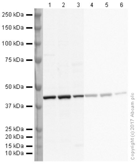
Western blot - Anti-ARMET/ARP antibody [EPR29115-74] (AB316935)
Exposure time : Lanes 1-3 : 180 seconds; Lanes 4-6 : 26 seconds.
All lanes:
Western blot - Anti-ARMET/ARP antibody [EPR29115-74] (ab316935) at 1/1000 dilution
Lane 1:
HeLa (human cervical adenocarcinoma epithelial cell) whole cell lysate at 20 µg
Lane 2:
SH-SY5Y (human neuroblastoma epithelial cell) whole cell lysate at 20 µg
Lane 3:
U-87 MG (human glioblastoma-astrocytoma epithelial cell) whole cell lysate at 20 µg
Lane 4:
NIH/3T3 (mouse embryonic fibroblast) whole cell lysate at 20 µg
Lane 5:
Neuro-2a (mouse neuroblastoma neuroblast) whole cell lysate at 20 µg
Lane 6:
C6 (rat glial tumor glial cell) whole cell lysate at 20 µg
Secondary
All lanes:
Western blot - Goat Anti-Rabbit IgG H&L (HRP) (<a href='/products/secondary-antibodies/goat-rabbit-igg-h-l-hrp-ab97051'>ab97051</a>) at 1/100000 dilution
Observed band size: 18 kDa
false

- WB
Supplier Data
Western blot - Anti-ARMET/ARP antibody [EPR29115-74] (AB316935)
Low expression : skeletal muscle (PMID : 31781038).
In Western blot, Anti-GAPDH antibody [EPR16891] - Loading Control (ab181602) staining at 1/200000 dilution.
All lanes:
Western blot - Anti-ARMET/ARP antibody [EPR29115-74] (ab316935) at 1/1000 dilution
Lane 1:
Human tonsil tissue lysate at 20 µg
Lane 2:
Human skeletal muscle tissue lysate at 20 µg
Secondary
All lanes:
Goat Anti-Rabbit IgG (HRP) with minimal cross-reactivity with human IgG at 1/2000 dilution
Observed band size: 18 kDa,36 kDa
false
Exposure time: 180s

- WB
Supplier Data
Western blot - Anti-ARMET/ARP antibody [EPR29115-74] (AB316935)
Low expression : brain, cerebral cortex, heart, skeletal muscle (PMID : 24587361; PMID : 31781038).
In Western blot, Anti-GAPDH antibody [EPR16891] - Loading Control (ab181602) staining at 1/200000 dilution.
All lanes:
Western blot - Anti-ARMET/ARP antibody [EPR29115-74] (ab316935) at 1/1000 dilution
Lane 1:
Mouse P5 brain tissue lysate at 20 µg
Lane 2:
Mouse brain tissue lysate at 20 µg
Lane 3:
Mouse cerebral cortex tissue lysate at 20 µg
Lane 4:
Mouse heart tissue lysate at 20 µg
Lane 5:
Mouse skeletal muscle tissue lysate at 20 µg
Lane 6:
Rat P5 brain tissue lysate at 20 µg
Lane 7:
Rat brain tissue lysate at 20 µg
Lane 8:
Rat heart tissue lysate at 20 µg
Lane 9:
Rat skeletal muscle tissue lysate at 20 µg
Secondary
All lanes:
Western blot - Goat Anti-Rabbit IgG H&L (HRP) (<a href='/products/secondary-antibodies/goat-rabbit-igg-h-l-hrp-ab97051'>ab97051</a>) at 1/100000 dilution
Observed band size: 18 kDa,36 kDa
false
Exposure time: 102s

- WB
Supplier Data
Western blot - Anti-ARMET/ARP antibody [EPR29115-74] (AB316935)
This antibody does not cross-react with mouse CDNF.
In Western blot, Anti-6X His tag® antibody [EPR20547] - ChIP Grade (ab213204) staining at 1/5000 dilution.
All lanes:
Western blot - Anti-ARMET/ARP antibody [EPR29115-74] (ab316935) at 1/1000 dilution
Lane 1:
His-tagged mouse MANF recombinant protein at 10 ng
Lane 2:
His-tagged mouse CDNF recombinant protein at 10 ng
Secondary
All lanes:
Western blot - Goat Anti-Rabbit IgG H&L (HRP) (<a href='/products/secondary-antibodies/goat-rabbit-igg-h-l-hrp-ab97051'>ab97051</a>) at 1/100000 dilution
Observed band size: 18 kDa
false
Exposure time: 26s
不同偶联物与剂型 (1)
-
Anti-ARMET/ARP antibody [EPR29115-74] - BSA and Azide free
搭配使用产品
Primary Antibodies
AB181602
Anti-GAPDH antibody [EPR16891] - Loading Control
RabMAb|Recombinant|Lab Essentials|20ul selling size

- 5
- 23
Primary Antibodies
AB8227
Anti-beta Actin antibody - Loading Control
BOND RX™ Validated|Lab Essentials
- 5
- 104
Primary Antibodies
AB300469
Anti-PSAP antibody [EPR25650-11]
BOND RX™ Validated|20ul selling size|RabMAb|Recombinant

- 0
- 0
Primary Antibodies
AB16667
BOND RX™ Validated|KO Validated|RabMAb|Advanced Validation|Recombinant|20ul selling size

- 5
- 133
反应性数据
产品详情
Patented technology
Our RabMAb® technology is a patented hybridoma-based technology for making rabbit monoclonal antibodies. For details on our patents, please refer to RabMAb® patents.
What are the advantages of a recombinant monoclonal antibody?
This product is a recombinant monoclonal antibody, which offers several advantages including:
- - High batch-to-batch consistency and reproducibility
- - Improved sensitivity and specificity
- - Long-term security of supply
- - Animal-free batch production
For more information, read more on recombinant antibodies.
性能和储存信息
形式
纯化工艺
存储溶液
运输条件
推荐的短期储存时间
推荐的短期储存条件
推荐的长期储存条件
分装信息
储存信息
补充信息
This supplementary information is collated from multiple sources and compiled automatically.
Biological function summary
ARMET plays a significant role in maintaining cellular homeostasis especially under stress conditions. It does not form a larger complex but functions as a standalone factor that aids in the proper folding of proteins within the endoplasmic reticulum. Additionally ARMET has protective effects on dopaminergic neurons which are critical for neuromolecular processes. This protein has connections to cellular survival pathways acting as a neurotrophic factor to support neuron maintenance and health.
Pathways
ARMET is an important player in the unfolded protein response (UPR) and the neuroprotective pathways. In these pathways ARMET interacts with other proteins involved in protein folding and stress responses such as BiP/GRP78. These interactions help prevent apoptosis and promote cell survival by ensuring that cells can cope with endoplasmic reticulum stress. The ability of ARMET to enhance neuronal survival links it to pathways important for neuroprotection.
产品实验方案
- Visit the General protocols
- Visit the Troubleshooting
靶点信息
文献 (1)
Recent publications for all applications. Explore the full list and refine your search
Scientific reports 15:476 PubMed39747250
2025
Applications
Unspecified application
Species
Unspecified reactive species
Abcam Product Promise
Please note: All products are 'FOR RESEARCH USE ONLY. NOT FOR USE IN DIAGNOSTIC OR THERAPEUTIC PROCEDURES'.
For licensing inquiries, please contact partnerships@abcam.com