重组Anti-AIRE抗体[EPR28045-139]
Anti-AIRE antibody [EPR28045-139]
- BOND RX™ Validated
- 20ul selling size
- Recombinant
- RabMAb
- KO Validated
- 了解详情
Be the first to review this product! Submit a review
|
(0 Publication)
Rabbit Recombinant Monoclonal AIRE antibody. Suitable for IHC-P, WB, IP and reacts with Mouse, Rat samples.
查看别名
Autoimmune regulator, Autoimmune polyendocrinopathy candidiasis ectodermal dystrophy protein homolog, APECED protein homolog, Aire

- IHC-P
Supplier Data
Immunohistochemistry (Formalin/PFA-fixed paraffin-embedded sections) - Anti-AIRE antibody [EPR28045-139] (AB315164)
Immunohistochemical analysis of paraffin-embedded Mouse thymus tissue labeling AIRE with ab315164 at 1/500 (1.034 ug/ml) dilution, followed by a ready to use LeicaDS9800 (Bond™ Polymer Refine Detection).
Nuclear staining on a few cells in the medulla of mouse thymus (PMID : 15492124; PMID : 28232470). The section was incubated with ab315164 for 30 mins at room temperature.
The immunostaining was performed on a Leica Biosystems BOND® RX instrument
Counterstained with Hematoxylin.
Secondary antibody only control : Secondary antibody is a ready to use LeicaDS9800 (Bond™ Polymer Refine Detection).
Heat mediated antigen retrieval was performed with Tris-EDTA buffer (pH 9.0, Epitope Retrieval Solution2) for 20 mins

- IHC-P
Supplier Data
Immunohistochemistry (Formalin/PFA-fixed paraffin-embedded sections) - Anti-AIRE antibody [EPR28045-139] (AB315164)
Immunohistochemical analysis of paraffin-embedded Rat thymus tissue labeling AIRE with ab315164 at 1/500 (1.034 ug/ml) dilution, followed by a ready to use LeicaDS9800 (Bond™ Polymer Refine Detection).
Nuclear staining on a few cells in the medulla of rat thymus. The section was incubated with ab315164 for 30 mins at room temperature.
The immunostaining was performed on a Leica Biosystems BOND® RX instrument
Counterstained with Hematoxylin.
Secondary antibody only control : Secondary antibody is a ready to use LeicaDS9800 (Bond™ Polymer Refine Detection).
Heat mediated antigen retrieval was performed with Tris-EDTA buffer (pH 9.0, Epitope Retrieval Solution2) for 20 mins
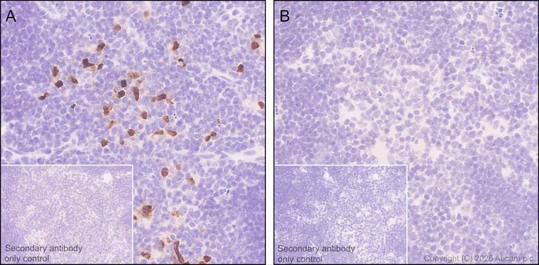
- IHC-P
Lab
Immunohistochemistry (Formalin/PFA-fixed paraffin-embedded sections) - Anti-AIRE antibody [EPR28045-139] (AB315164)
Immunohistochemical analysis of paraffin-embedded (A) Thymus tissue from wild-type C57BL/6JGpt mice (B) Thymus tissue from Aire knockout mice labelling AIRE with ab315164 at 1/500 (1.034 ug/ml) followed by a ready to use LeicaDS9800 (Bond™ Polymer Refine Detection).
Positive staining on (A) Thymus tissue from wild-type C57BL/6JGpt mice and no staining on (B) Thymus tissue from Aire knockout mice.
The primary antibody was incubated for 30 mins at room temperature.
The immunostaining was performed on a Leica Biosystems BOND® RX instrument.
The tissue samples were kindly provided by GemPharmatech C57BL/6JGpt wildtype mice and Aire-KO homozygous mice (Strain ID : T017083).
Heat mediated antigen retrieval was performed with Tris-EDTA buffer (pH 9.0, Epitope Retrieval Solution2) for 20 mins

- IHC-P
Supplier Data
Immunohistochemistry (Formalin/PFA-fixed paraffin-embedded sections) - Anti-AIRE antibody [EPR28045-139] (AB315164)
Immunohistochemical analysis of paraffin-embedded Rat cerebrum tissue labeling AIRE with ab315164 at 1/500 (1.034 ug/ml) dilution, followed by a ready to use LeicaDS9800 (Bond™ Polymer Refine Detection).
Negative control : no staining on rat cerebrum. The section was incubated with ab315164 for 30 mins at room temperature.
The immunostaining was performed on a Leica Biosystems BOND® RX instrument
Counterstained with Hematoxylin.
Secondary antibody only control : Secondary antibody is a ready to use LeicaDS9800 (Bond™ Polymer Refine Detection).
Heat mediated antigen retrieval was performed with Tris-EDTA buffer (pH 9.0, Epitope Retrieval Solution2) for 20 mins

- IHC-P
Supplier Data
Immunohistochemistry (Formalin/PFA-fixed paraffin-embedded sections) - Anti-AIRE antibody [EPR28045-139] (AB315164)
Immunohistochemical analysis of paraffin-embedded Mouse cerebrum tissue labeling AIRE with ab315164 at 1/500 (1.034 ug/ml) dilution, followed by a ready to use LeicaDS9800 (Bond™ Polymer Refine Detection).
Negative control : no staining on mouse cerebrum (PMID : 10925280). The section was incubated with ab315164 for 30 mins at room temperature.
The immunostaining was performed on a Leica Biosystems BOND® RX instrument
Counterstained with Hematoxylin.
Secondary antibody only control : Secondary antibody is a ready to use LeicaDS9800 (Bond™ Polymer Refine Detection).
Heat mediated antigen retrieval was performed with Tris-EDTA buffer (pH 9.0, Epitope Retrieval Solution2) for 20 mins

- IHC-P
Supplier Data
Immunohistochemistry (Formalin/PFA-fixed paraffin-embedded sections) - Anti-AIRE antibody [EPR28045-139] (AB315164)
Immunohistochemical analysis of paraffin-embedded Rat liver tissue labeling AIRE with ab315164 at 1/500 (1.034 ug/ml) dilution, followed by a ready to use LeicaDS9800 (Bond™ Polymer Refine Detection).
Negative control : no staining on rat liver. The section was incubated with ab315164 for 30 mins at room temperature.
The immunostaining was performed on a Leica Biosystems BOND® RX instrument
Counterstained with Hematoxylin.
Secondary antibody only control : Secondary antibody is a ready to use LeicaDS9800 (Bond™ Polymer Refine Detection).
Heat mediated antigen retrieval was performed with Tris-EDTA buffer (pH 9.0, Epitope Retrieval Solution2) for 20 mins

- IHC-P
Supplier Data
Immunohistochemistry (Formalin/PFA-fixed paraffin-embedded sections) - Anti-AIRE antibody [EPR28045-139] (AB315164)
Immunohistochemical analysis of paraffin-embedded Mouse liver tissue labeling AIRE with ab315164 at 1/500 (1.034 ug/ml) dilution, followed by a ready to use LeicaDS9800 (Bond™ Polymer Refine Detection).
Negative control : no staining on mouse liver (PMID : 10925280). The section was incubated with ab315164 for 30 mins at room temperature.
The immunostaining was performed on a Leica Biosystems BOND® RX instrument
Counterstained with Hematoxylin.
Secondary antibody only control : Secondary antibody is a ready to use LeicaDS9800 (Bond™ Polymer Refine Detection).
Heat mediated antigen retrieval was performed with Tris-EDTA buffer (pH 9.0, Epitope Retrieval Solution2) for 20 mins

- IP
Supplier Data
Immunoprecipitation - Anti-AIRE antibody [EPR28045-139] (AB315164)
AIRE was immunoprecipitated from 0.35 mg Mouse thymus tissue lysate with ab315164 at 1/30 dilution (2ug in 0.35mg lysates). Western blot was performed on the immunoprecipitate using ab315164 at 1/1000 dilution. VeriBlot for IP secondary antibody(HRP)(ab131366) was used at 1/5000 dilution.
Lane 1 : Mouse thymus tissue lysate
Lane 2 : ab315164 IP in Mouse thymus tissue lysate
Lane 3 : Rabbit monoclonal IgG (ab172730) instead of ab315164 in Mouse thymus tissue lysate
All lanes:
Immunoprecipitation - Anti-AIRE antibody [EPR28045-139] (ab315164) at 1/30 dilution
All lanes:
Mouse thymus tissue lysate
Secondary
All lanes:
Immunoprecipitation - VeriBlot for IP Detection Reagent (HRP) (<a href='/products/reagents/veriblot-for-ip-detection-reagent-hrp-ab131366'>ab131366</a>) at 1/5000 dilution
false
Exposure time: 180s

- WB
Supplier Data
Western blot - Anti-AIRE antibody [EPR28045-139] (AB315164)
Negative control : mouse kidney (PMID : 18214467, PMID : 10925280), mouse liver (PMID : 18214467, PMID : 10925280), mouse skin (PMID : 18214467, PMID : 10925280).
In Western blot, Anti-GAPDH antibody [EPR16891] - Loading Control (ab181602) staining at 1/200000 dilution.
All lanes:
Western blot - Anti-AIRE antibody [EPR28045-139] (ab315164) at 1/1000 dilution
Lane 1:
Mouse thymus tissue lysate at 50 µg
Lane 2:
Mouse kidney tissue lysate at 50 µg
Lane 3:
Mouse liver tissue lysate at 50 µg
Lane 4:
Mouse skin tissue lysate at 50 µg
Secondary
All lanes:
Goat Anti-Rabbit IgG (HRP) with minimal cross-reactivity with human IgG at 1/2000 dilution
Observed band size: 59 kDa,36 kDa
false
Exposure time: 26s
不同偶联物与剂型 (3)
-
Anti-AIRE antibody [EPR28045-139] - BSA and Azide free
-
519 Alexa Fluor® 488
Alexa Fluor® 488 Anti-AIRE antibody [EPR28045-139]
-
665 Alexa Fluor® 647
Alexa Fluor® 647 Anti-AIRE antibody [EPR28045-139]
搭配使用产品
Primary Antibodies
AB53280
Anti-Cytokeratin 8 antibody [EP1628Y] - Cytoskeleton Marker
KO Validated|RabMAb|Recombinant|Lab Essentials|20ul selling size

- 5
- 9
Primary Antibodies
AB119695
Anti-Cytokeratin 14 antibody [SP53]
BOND RX™ Validated|20ul selling size|RabMAb|Advanced Validation|Recombinant|KO Validated

- 5
- 6
Primary Antibodies
AB183734
RabMAb|Recombinant|Lab Essentials|20ul selling size

- 5
- 24
Primary Antibodies
AB281583
BOND RX™ Validated|RabMAb|Recombinant|Lab Essentials|20ul selling size

- 5
- 5
反应性数据
产品详情
Patented technology
Our RabMAb® technology is a patented hybridoma-based technology for making rabbit monoclonal antibodies. For details on our patents, please refer to RabMAb® patents.
What are the advantages of a recombinant monoclonal antibody?
This product is a recombinant monoclonal antibody, which offers several advantages including:
- - High batch-to-batch consistency and reproducibility
- - Improved sensitivity and specificity
- - Long-term security of supply
- - Animal-free batch production
For more information, read more on recombinant antibodies.
性能和储存信息
形式
纯化工艺
存储溶液
运输条件
推荐的短期储存时间
推荐的短期储存条件
推荐的长期储存条件
分装信息
储存信息
补充信息
This supplementary information is collated from multiple sources and compiled automatically.
Biological function summary
AIRE functions to maintain central tolerance in the immune system. It acts as a part of a transcriptional complex that regulates genes involved in immune tolerance. This complex controls the expression of tissue-specific antigens (TSAs) important for "educating" T cells in the thymus to distinguish between self and non-self antigens. By controlling this mechanism AIRE helps to lower the risk of the body attacking its own tissues.
Pathways
AIRE contributes significantly to the pathway of immune tolerance and T cell selection within the thymus. It interacts with several other proteins involved in these pathways including the tumor necrosis factor receptor superfamily member RANK influencing the maturation of mTECs. Additionally AIRE interacts with proteins like CHD4 and a range of histone-modifying enzymes to modulate gene expression more broadly within the context of epigenetic regulation.
产品实验方案
- Visit the General protocols
- Visit the Troubleshooting
靶点信息
Abcam Product Promise
Please note: All products are 'FOR RESEARCH USE ONLY. NOT FOR USE IN DIAGNOSTIC OR THERAPEUTIC PROCEDURES'.
For licensing inquiries, please contact partnerships@abcam.com