Anti-Calnexin抗体- ER Marker (ab22595)
")
Key features and details
- Rabbit polyclonal to Calnexin - ER Marker
- Suitable for: WB, ICC/IF, IP
- Knockout validated
- Reacts with: Mouse, Rat, Human
- Isotype: IgG
选择批间可重复性更高的重组抗体


- 研究可靠 —— 各批次间结果一致且可重复
- 长期批量供应 —— 采用重组技术,可实现快速生产
- 首次实验即可成功 —— 经过大量验证确认了特异性
- 符合伦理标准 —— 产品不含动物成分
概述
-
产品名称
Anti-Calnexin抗体- ER Marker
参阅全部 Calnexin 一抗 -
描述
兔多克隆抗体to Calnexin - ER Marker -
宿主
Rabbit -
特异性
Recognizes ER membrane, mitochondria and cis-Golgi -
经测试应用
适用于: WB, ICC/IF, IPmore details -
种属反应性
与反应: Mouse, Rat, Human
预测可用于: Dog, Common marmoset
-
免疫原
Synthetic peptide corresponding to Human Calnexin aa 550 to the C-terminus (C terminal) conjugated to keyhole limpet haemocyanin.
(Peptide available asab23379) -
常规说明
The Life Science industry has been in the grips of a reproducibility crisis for a number of years. Abcam is leading the way in addressing this with our range of recombinant monoclonal antibodies and knockout edited cell lines for gold-standard validation. Please check that this product meets your needs before purchasing.
If you have any questions, special requirements or concerns, please send us an inquiry and/or contact our Support team ahead of purchase. Recommended alternatives for this product can be found below, along with publications, customer reviews and Q&As
性能
-
形式
Liquid -
存放说明
Shipped at 4°C. Store at +4°C short term (1-2 weeks). Upon delivery aliquot. Store at -20°C or -80°C. Avoid freeze / thaw cycle. Store In the Dark. -
存储溶液
pH: 7.40
Preservative: 0.02% Sodium azide
Constituent: PBS
Batches of this product that have a concentration < 1mg/ml may have BSA added as a stabilising agent. If you would like information about the formulation of a specific lot, please contact our scientific support team who will be happy to help. -
 Concentration information loading...
 -
纯度
Immunogen affinity purified -
克隆
多克隆 -
同种型
IgG -
研究领域
相关产品
-
Compatible Secondaries
-
Isotype control
-
Recombinant Protein
-
Related Products
应用
The Abpromise guarantee
Abpromise™承诺保证使用ab22595于以下的经测试应用
“应用说明”部分 下显示的仅为推荐的起始稀释度;实际最佳的稀释度/浓度应由使用者检定。
| 应用 | Ab评论 | 说明 |
|---|---|---|
| WB | (13) |
Use a concentration of 1 µg/ml. Detects a band of approximately 75 kDa (predicted molecular weight: 90 kDa).
|
| ICC/IF | (7) |
Use a concentration of 1 µg/ml.
|
| IP |
Use at an assay dependent concentration.
|
| 说明 |
|---|
|
WB
Use a concentration of 1 µg/ml. Detects a band of approximately 75 kDa (predicted molecular weight: 90 kDa). |
|
ICC/IF
Use a concentration of 1 µg/ml. |
|
IP
Use at an assay dependent concentration. |
靶标
-
功能
Calcium-binding protein that interacts with newly synthesized glycoproteins in the endoplasmic reticulum. It may act in assisting protein assembly and/or in the retention within the ER of unassembled protein subunits. It seems to play a major role in the quality control apparatus of the ER by the retention of incorrectly folded proteins. -
序列相似性
Belongs to the calreticulin family. -
细胞定位
Endoplasmic reticulum membrane. Melanosome. Identified by mass spectrometry in melanosome fractions from stage I to stage IV. - Information by UniProt
-
数据库链接
- Entrez Gene: 403908 Dog
- Entrez Gene: 821 Human
- Entrez Gene: 12330 Mouse
- Entrez Gene: 29144 Rat
- Omim: 114217 Human
- SwissProt: P24643 Dog
- SwissProt: P27824 Human
- SwissProt: P35564 Mouse
see all -
别名
- Calnexin antibody
- CALX_HUMAN antibody
- CANX antibody
see all
图片
-
Western blot - Anti-Calnexin antibody - ER Marker (ab22595)Dombert et al PLoS One. 2014 Oct 22;9(10):e110846. doi: 10.1371/journal.pone.0110846. eCollection 2014. Fig 1. Reproduced under the Creative Commons license http://creativecommons.org/licenses/by/4.0/
Subcellular distribution of Smn and hnRNP R in isolated mouse embryonic motoneurons.
Lentiviral knockdown of hnRNP R led to a dose-dependent reduction of hnRNP R levels. Calnexin and Smn protein were not altered significantly.
Primary motoneurons or E18 spinal cord tissue, respectively, were lysed with cytosolic and nuclear fractionation buffer, solubilized in Laemmli buffer and boiled for 10 minutes at 99°C. Proteins were then subjected to SDS-PAGE, blotted onto PVDF membrane, incubated with the corresponding antibodies, including ab22595.
-
 Western blot - Anti-Calnexin antibody - ER Marker (ab22595)
Lane 1: Wild type HAP1 whole cell lysate (20 µg)
Lane 2: empty lane
Lane 3: CANX knockout HAP1 whole cell lysate (20 µg)
Lane 4: empty lane
Lanes 1 - 4: Merged signal (red and green). Green - ab22595 observed at 80 kDa. Red - loading control, ab8245, observed at 37 kDa.
ab22595 was shown to specifically react with CANX (Calnexin) in wildtype cells as signal was lost in CANX (Calnexin) knockout cells. Wild-type and eCANX (Calnexin) knockout samples were subjected to SDS-PAGE. ab22595 and ab8245 (Mouse anti GAPDH loading control) were incubated overnight at 4°C at 1 dilution and 1/10,000 dilution respectively.Blots were developed with Goat anti-Rabbit IgG H&L (IRDye® 800CW) preabsorbed ab216773 and Goat anti-Mouse IgG H&L (IRDye® 680RD) preabsorbed ab216776 secondary antibodies at 1/10,000 dilution for 1 hour at room temperature before imaging.
-
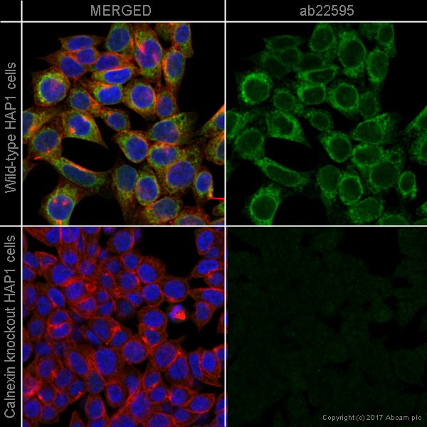 Immunocytochemistry/ Immunofluorescence - Anti-Calnexin antibody - ER Marker (ab22595)
ab22595 staining Calnexin in wild-type HAP1 cells (top panel) and CANX knockout HAP1 cells (bottom panel).
The cells were fixed with 4% formaldehyde for 10 minutes, permeabilized with 0.1% Triton X-100 for 5 minutes and then blocked with 1% BSA/10% normal goat serum/0.3M glycine in 0.1% PBS-Tween for 1 hour. The cells were then incubated with ab22595 at 1 μg/ml and ab195889 at 1/250 dilution (shown in pseudocolour red) overnight at +4°C, followed by a further incubation at room temperature for 1 hour with a goat secondary antibody to Rabbit IgG (Alexa Fluor® 488) (ab150081) at 2 μg/ml (shown in green).
Nuclear DNA was labeled in blue with DAPI.
Image was taken with a confocal microscope (Leica-Microsystems, TCS SP8).
-
 Immunocytochemistry/ Immunofluorescence - Anti-Calnexin antibody - ER Marker (ab22595)
ab22595 staining Calnexin in HeLa (Human epithelial cell line from cervix adenocarcinoma) cells.
The cells were fixed with 100% methanol for 5 minutes, permeabilized with 0.1% Triton X-100 for 5 minutes and then blocked with 1% BSA/10% normal goat serum/0.3M glycine in 0.1% PBS-Tween for 1 hour. The cells were then incubated with ab22595 at 1 μg/ml and ab7291 at 1 μg/ml overnight at +4°C, followed by a further incubation at room temperature for 1 hour with a goat secondary antibody to Rabbit IgG (Alexa Fluor® 488) (ab150081) at 2 μg/ml (shown in green) and a goat secondary antibody to Mouse IgG (Alexa Fluor® 594) (ab150120).
Nuclear DNA was labeled in blue with DAPI.
Image was taken with a confocal microscope (Leica-Microsystems, TCS SP8).
-
 Western blot - Anti-Calnexin antibody - ER Marker (ab22595)
Lane 1: Wild-type HAP1 cell lysate (20 µg)
Lane 2: Calnexin knockout HAP1 cell lysate (20 µg)Lanes 1 - 2: Merged signal (red and green). Green - ab22595 observed at 80 kDa. Red - loading control, ab8245, observed at 37 kDa.
This western blot image is a comparison between ab22595 and a competitor's top cited rabbit polyclonal antibody.
-
 Western blot - Anti-Calnexin antibody - ER Marker (ab22595)All lanes : Anti-Calnexin antibody - ER Marker (ab22595) at 1/250 dilution
Lane 1 :NIH/3T3 whole cell lysate (ab7179)
Lane 2 : MEF1 (Mouse embryonic fibroblast cell line) Whole Cell Lysate (ab46770)
Lane 3 : Brain (Mouse) Tissue Lysate (ab27253)
Lane 4 : Liver (Mouse) Tissue Lysate (ab7935)
Lane 5 : Heart (Mouse) Tissue Lysate (ab27255)
Lane 6 : Kidney (Mouse) Tissue Lysate (ab27254)
Lane 7 :Mouse pancreas tissue lysate - total protein (ab29363)
Lane 8 : Testis (Mouse) Tissue Lysate - normal tissue (ab4027)
Lane 9 :Mouse skeletal muscle tissue lysate - total protein (ab29711)
Lane 10 : Spinal Cord (Mouse) Tissue Lysate (ab50253)
Lane 11 : Ovary (Mouse) Tissue Lysate (ab35808)
Lane 12 : PC-12 (Rat adrenal pheochromocytoma cell line) Whole Cell Lysate (ab50957)
Lane 13 : Brain (Rat) Tissue Lysate (ab7942)
Lane 14 : Liver (Rat) Tissue Lysate (ab27256)
Lane 15 : Heart (Rat) Tissue Lysate (ab7940)
Lane 16 : Kidney (Rat) Whole Cell Lysate - normal tissue (ab29480)
Lysates/proteins at 10 µg per lane.
Secondary
All lanes : IRDye 680 Conjugated Goat Anti-Rabbit IgG (H+L) at 1/10000 dilution
Performed under reducing conditions.
Predicted band size: 90 kDa
Observed band size: 80 kDa why is the actual band size different from the predicted? -
 Western blot - Anti-Calnexin antibody - ER Marker (ab22595)All lanes : Anti-Calnexin antibody - ER Marker (ab22595) at 1 µg/ml
Lane 1 : HeLa (Human epithelial carcinoma cell line) whole cell lysate
Lane 2 : U-2 OS (Human bone osteosarcoma epithelial cell line) whole cell lysate
Lane 3 : MCF7 (Human breast adenocarcinoma cell line) whole cell lysate
Lane 4 : HeLa whole cell lysate with Human Calnexin peptide (ab23379) at 1 µg/ml
Lane 5 : U-2 OS whole cell lysate with Human Calnexin peptide (ab23379) at 1 µg/ml
Lane 6 : MCF7 whole cell lysate with Human Calnexin peptide (ab23379) at 1 µg/ml
Lysates/proteins at 20 µg per lane.
Secondary
All lanes : Goat polyclonal to Rabbit IgG (Alexa Fluor® 680) at 1/10000 dilution
Performed under reducing conditions.
Predicted band size: 90 kDa
Observed band size: 75 kDa why is the actual band size different from the predicted?Recent batches of ab22595 (AP217379 and AP151845) detect a band of ~ 75 kDa in HeLa, U-2 OS and MCF7 lysates. This band is completely blocked by the immunizing peptide so we believe this represents Calnexin. Moreoever, a band of the same size is detected by other Calnexin antibodies tested.
-
 Immunoprecipitation - Anti-Calnexin antibody - ER Marker (ab22595)
Calnexin was immunoprecipitated using 0.5 mg HeLa (Human epithelial cell line from cervix adenocarcinoma) whole cell extract, 5 µg of Rabbit polyclonal to Calnexin - ER membrane marker and 50 µl of protein G magnetic beads (+).
No antibody was added to the control (-).
The antibody was incubated under agitation with Protein G beads for 10 minutes, HeLa whole cell extract lysate diluted in RIPA buffer was added to each sample and incubated for a further 10 minutes under agitation.
Proteins were eluted by addition of 40 µl SDS loading buffer and incubated for 10 minutes at 70oC; 10 µl of each sample was separated on a SDS PAGE gel, transferred to a nitrocellulose membrane, blocked with 5% BSA and probed with ab22595.
Secondary: Goat polyclonal to mouse IgG light chain specific (HRP) at 1/5000 dilution.
Band: 80kDa: Calnexin - ER membrane marker.
实验方案
数据表及文件
-
SDS download
-
Datasheet download
文献 (378)
ab22595 被引用在 378 文献中.
- Niki Y et al. S-Palmitoylation of Tyrosinase at Cysteine500 Regulates Melanogenesis. J Invest Dermatol 143:317-327.e6 (2023). PubMed: 36063887
- Wang C et al. A light-activated magnetic bead strategy utilized in spatio-temporal controllable exosomes isolation. Front Bioeng Biotechnol 10:1006374 (2022). PubMed: 36147530
- Chen X et al. Screening of plasma exosomal lncRNAs to identify potential biomarkers for obstructive sleep apnea. Ann Transl Med 10:936 (2022). PubMed: 36172105
- Hu Q et al. Emodin attenuates severe acute pancreatitis-associated acute lung injury by suppressing pancreatic exosome-mediated alveolar macrophage activation. Acta Pharm Sin B 12:3986-4003 (2022). PubMed: 36213542
- Sadovska L et al. Effects of urinary extracellular vesicles from prostate cancer patients on the transcriptomes of cancer-associated and normal fibroblasts. BMC Cancer 22:1055 (2022). PubMed: 36224527