Anti-VE Cadherin抗体- Intercellular Junction Marker (ab33168)
")
Key features and details
- Rabbit polyclonal to VE Cadherin - Intercellular Junction Marker
- Suitable for: ICC/IF, WB
- Reacts with: Mouse, Human
- Isotype: IgG
选择批间可重复性更高的重组抗体


- 研究可靠 —— 各批次间结果一致且可重复
- 长期批量供应 —— 采用重组技术,可实现快速生产
- 首次实验即可成功 —— 经过大量验证确认了特异性
- 符合伦理标准 —— 产品不含动物成分
概述
-
产品名称
Anti-VE Cadherin抗体- Intercellular Junction Marker
参阅全部 VE Cadherin 一抗 -
描述
兔多克隆抗体to VE Cadherin - Intercellular Junction Marker -
宿主
Rabbit -
经测试应用
适用于: ICC/IF, WBmore details -
种属反应性
与反应: Mouse, Human
预测可用于: Chicken, Cow, Pig
-
免疫原
Synthetic peptide corresponding to Human VE Cadherin aa 750 to the C-terminus conjugated to keyhole limpet haemocyanin.
(Peptide available asab27462) -
阳性对照
- ICC/IF: HUVEC cells. WB: HUVEC cell lysate and Mouse lung tissue lysate.
-
常规说明
The Life Science industry has been in the grips of a reproducibility crisis for a number of years. Abcam is leading the way in addressing this with our range of recombinant monoclonal antibodies and knockout edited cell lines for gold-standard validation. Please check that this product meets your needs before purchasing.
If you have any questions, special requirements or concerns, please send us an inquiry and/or contact our Support team ahead of purchase. Recommended alternatives for this product can be found below, along with publications, customer reviews and Q&As
性能
-
形式
Liquid -
存放说明
Shipped at 4°C. Store at +4°C short term (1-2 weeks). Upon delivery aliquot. Store at -20°C or -80°C. Avoid freeze / thaw cycle. -
存储溶液
pH: 7.40
Preservative: 0.02% Sodium azide
Constituent: PBS
1x PBS
Batches which are <1mg/ml will contain 1% BSA, batches at 1mg/ml will not. -
 Concentration information loading...
 -
纯度
Immunogen affinity purified -
克隆
多克隆 -
同种型
IgG -
研究领域
相关产品
-
Compatible Secondaries
-
Conjugation kits
-
Isotype control
-
Recombinant Protein
-
Related Products
应用
The Abpromise guarantee
Abpromise™承诺保证使用ab33168于以下的经测试应用
“应用说明”部分 下显示的仅为推荐的起始稀释度;实际最佳的稀释度/浓度应由使用者检定。
| 应用 | Ab评论 | 说明 |
|---|---|---|
| ICC/IF | (18) |
Use a concentration of 0.1 - 1 µg/ml.
Abcam recommends using this product with confluent cells. |
| WB | (9) |
Use a concentration of 1 µg/ml. Detects a band of approximately 115,117,120 kDa (predicted molecular weight: 88 kDa).
Abcam recommends using BSA blocking with this product. Milk blocking will give a greatly reduced signal strength in WB. |
| 说明 |
|---|
|
ICC/IF
Use a concentration of 0.1 - 1 µg/ml. Abcam recommends using this product with confluent cells. |
|
WB
Use a concentration of 1 µg/ml. Detects a band of approximately 115,117,120 kDa (predicted molecular weight: 88 kDa). Abcam recommends using BSA blocking with this product. Milk blocking will give a greatly reduced signal strength in WB. |
靶标
-
功能
Cadherins are calcium dependent cell adhesion proteins. They preferentially interact with themselves in a homophilic manner in connecting cells; cadherins may thus contribute to the sorting of heterogeneous cell types. This cadherin may play a important role in endothelial cell biology through control of the cohesion and organization of the intercellular junctions. It associates with alpha-catenin forming a link to the cytoskeleton. -
组织特异性
Endothelial tissues and brain. -
序列相似性
Contains 5 cadherin domains. -
翻译后修饰
Phosphorylated on tyrosine residues by KDR/VEGFR-2. Dephosphorylated by PTPRB. -
细胞定位
Cell junction. Cell membrane. Found at cell-cell boundaries and probably at cell-matrix boundaries. - Information by UniProt
-
数据库链接
- Entrez Gene: 1003 Human
- Entrez Gene: 12562 Mouse
- Omim: 601120 Human
- SwissProt: P33151 Human
- SwissProt: P55284 Mouse
- Unigene: 76206 Human
- Unigene: 21767 Mouse
-
别名
- 7B 4 antibody
- 7B4 antibody
- 7B4 antigen antibody
see all
图片
-
Anti-VE Cadherin antibody - Intercellular Junction Marker (ab33168)This image is courtesy of an Abreview submitted by Simon Shen
-
 Immunocytochemistry/ Immunofluorescence - Anti-VE Cadherin antibody - Intercellular Junction Marker (ab33168)
ab33168 staining VE Cadherin in HUV-EC cells. The cells were fixed with 100% methanol (5 min), permeabilized with 0.1% PBS-Tween for 5 minutes and then blocked with 1% BSA/10% normal goat serum/0.3M glycine in 0.1% PBS-Tween for 1h. The cells were then incubated overnight at 4°C with ab33168 at 1µg/ml and ab7291, Mouse monoclonal [DM1A] to alpha Tubulin - Loading Control. Cells were then incubated with ab150081, Goat polyclonal Secondary Antibody to Rabbit IgG - H&L (Alexa Fluor® 488), pre-adsorbed at 1/1000 dilution (shown in green) and ab150120, Goat polyclonal Secondary Antibody to Mouse IgG - H&L (Alexa Fluor® 594), pre-adsorbed at 1/1000 dilution (shown in pseudocolour red). Nuclear DNA was labelled with DAPI (shown in blue).
Image was acquired with a high-content analyser (Operetta CLS, Perkin Elmer) and a maximum intensity projection of confocal sections is shown.
-
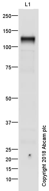
 Western blot - Anti-VE Cadherin antibody - Intercellular Junction Marker (ab33168)All lanes : Anti-VE Cadherin antibody - Intercellular Junction Marker (ab33168) at 1 µg/ml
Lane 1 : HUVEC (Human Umbilical Vein Endothelial Cell) Whole Cell Lysate
Lane 2 : Mouse lung tissue lysate
Lysates/proteins at 10 µg per lane.
Secondary
All lanes : Goat polyclonal to Rabbit IgG - H&L - Pre-Adsorbed (HRP) at 1/50000 dilution
Predicted band size: 88 kDa
Observed band size: 120 kDa why is the actual band size different from the predicted?
Additional bands at: 70 kDa (possible non-specific binding)
Exposure time: 1 minuteGel type: MOPS
Blocking buffer: 2% BSA -
 Western blot - Anti-VE Cadherin antibody - Intercellular Junction Marker (ab33168)Anti-VE Cadherin antibody - Intercellular Junction Marker (ab33168) at 1 µg/ml + HUVEC Cell Lysate at 10 µg
Secondary
Goat polyclonal to Rabbit IgG - H&L - Pre-Adsorbed (HRP) at 1/50000 dilution
Developed using the ECL technique.
Performed under reducing conditions.
Predicted band size: 88 kDa
Observed band size: 120 kDa why is the actual band size different from the predicted?
Exposure time: 1 minuteThis blot was produced using a 4-12% Bis-tris gel under the MOPS buffer system. The gel was run at 200V for 50 minutes before being transferred onto a Nitrocellulose membrane at 30V for 70 minutes. The membrane was then blocked for an hour using 2% Bovine Serum Albumin before being incubated with ab33168 overnight at 4°C. Antibody binding was detected using an anti-rabbit antibody conjugated to HRP, and visualised using ECL development solution ab133406.
The band we observe at 115 kDa is believed to be the glycosylated form of the protein.
-
 Western blot - Anti-VE Cadherin antibody - Intercellular Junction Marker (ab33168)Anti-VE Cadherin antibody - Intercellular Junction Marker (ab33168) at 1 µg/ml + HUVEC Cell Lysate at 10 µg
Secondary
Goat polyclonal to Rabbit IgG - H&L - Pre-Adsorbed (HRP) at 1/50000 dilution
Developed using the ECL technique.
Performed under reducing conditions.
Predicted band size: 88 kDa
Observed band size: 120 kDa why is the actual band size different from the predicted?
Additional bands at: 55 kDa (possible non-specific binding)
Exposure time: 1 minuteThis blot was produced using a 4-12% Bis-tris gel under the MOPS buffer system. The gel was run at 200V for 50 minutes before being transferred onto a Nitrocellulose membrane at 30V for 70 minutes. The membrane was then blocked for an hour using 2% Bovine Serum Albumin before being incubated with ab33168 overnight at 4°C. Antibody binding was detected using an anti-rabbit antibody conjugated to HRP, and visualised using ECL development solution ab133406.
The band we observe at 115 kDa is believed to be the glycosylated form of the protein.
-
 Immunocytochemistry/ Immunofluorescence - Anti-VE Cadherin antibody - Intercellular Junction Marker (ab33168)This image is courtesy of Stephen Yarwood, Inst Mol, Cell and Sys Bio, United KingdomICC/IF image of VE-Cadherin staining on HUVEC cells using ab33168. The cells were incubated with the primary antibody (ab33168) and the secondary was FITC conjugated anti-rabbit used at 1:400. The cells were incubated with only the secondary antibody as a negative control.
 -
 Immunocytochemistry/ Immunofluorescence - Anti-VE Cadherin antibody - Intercellular Junction Marker (ab33168)This image is courtesy of Ana Kasirer-Friede, Univ California-San Diego, Dept. Of Medicine, United States
ICC/IF image of VE Cadherin stained HUVEC cells. The cells were incubated with the antibody ab33168 at 1/150 (Green). The cells were also stained with Rhodamine phalloidin (Red).
-
 Western blot - Anti-VE Cadherin antibody - Intercellular Junction Marker (ab33168)Anti-VE Cadherin antibody - Intercellular Junction Marker (ab33168) at 1 µg/ml + HUVEC Cell Lysate at 10 µg
Secondary
Goat polyclonal to Rabbit IgG - H&L - Pre-Adsorbed (HRP) at 1/50000 dilution
Developed using the ECL technique.
Performed under reducing conditions.
Predicted band size: 88 kDa
Observed band size: 115,117 kDa why is the actual band size different from the predicted?
Additional bands at: 45 kDa (possible non-specific binding)
Exposure time: 1 minuteThe observed band for Cadherin 5 has a higher molecular weight of 115kDa due to glycosylation of the protein.
The immunogen used to raise this antibody has 89% homology with Cadherin 18, 88kDa , which we believe is the additional observed band at 117kDa, again due to glycosylation of the protein.
-
 Immunocytochemistry/ Immunofluorescence - Anti-VE Cadherin antibody - Intercellular Junction Marker (ab33168)This image is courtesy of an Abreview submitted by Kara Shumansky
ab33168 staining VE Cadherin in the endothelial cell line from Human liver by ICC/IF (Immunocytochemistry/immunofluorescence). Cells were fixed with Paraformaldehyde. Samples were incubated with primary antibody (1/100 in PBS + 2.5% BSA + 0.1% triton) for 1 hour at 37°C. Alexa Fluor 594 Chicken anti-Rabbit IgG (H+L) Cross-Adsorbed Secon was used as the secondary antibody at 4 µg/ml.
实验方案
数据表及文件
-
SDS download
-
Datasheet download
文献 (312)
ab33168 被引用在 312 文献中.
- Dolmatova EV et al. Endothelial Poldip2 regulates sepsis-induced lung injury via Rho pathway activation. Cardiovasc Res 118:2506-2518 (2022). PubMed: 34528082
- Brinks J et al. The Cortisol Response of Male and Female Choroidal Endothelial Cells: Implications for Central Serous Chorioretinopathy. J Clin Endocrinol Metab 107:512-524 (2022). PubMed: 34546342
- Li B et al. RNA N6-methyladenosine modulates endothelial atherogenic responses to disturbed flow in mice. Elife 11:N/A (2022). PubMed: 35001873
- Harati R et al. miR-27a-3p regulates expression of intercellular junctions at the brain endothelium and controls the endothelial barrier permeability. PLoS One 17:e0262152 (2022). PubMed: 35025943
- Jiang ZZ et al. Neutrophil extracellular traps induce tumor metastasis through dual effects on cancer and endothelial cells. Oncoimmunology 11:2052418 (2022). PubMed: 35309732